[Menu] [Previous] [Next]
TUTORIAL: Radioisotope Production
Production of Radioisotopes
Use the "Menu" button to jump to the Let's Play PET Main Menu or click on the [Next] and [Previous] buttons to proceed sequentially through the topics and tutorials. Or, you can return to the Department of Molecular and Medical Pharmacology's Home Page.Contents:
Topics:

Click on image above to view full-size image.
Shown here are the three major components of a radioisotope delivery system (RDS). The cyclotron is a particle accelerator used for the production of radioisotopes. These radioisotopes are then attached to biologically relevant molecules using a biosynthesizer unit. The entire process is controlled by a personal computer.
Cyclotron Operation
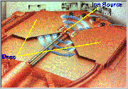
Click on image above to view full-size image.
A cyclotron consists of a pair of hollow, semicircular metal electrodes (called "dees" because of their shape), positioned between the poles of a large electromagnet (not shown). The dees are separated from one another by a narrow gap. Near the center of the dees is an ion source (typically an electrical arc device in a gas) that is used to generate charged particles.

Click on image above to view full-size image.
During operation, particles, such as the hydrogen ion depicted here, are generated in bursts by the ion source. A filament located in the ion source assembly creates a negative charge on the hydrogen ions through the addition of two electrons to the hydrogen (the electrons are depicted in yellow in the following screens).
As the negative ions enter the vacuum tank, they gain energy due to a high-frequency alternating electric field induced on the dees. As the negative ions flow from the ion source, they are exposed to this electric field as well as a strong magnetic field generated by two magnet poles, one above and one below the vacuum tank. Because these are charged particles in a magnetic field, the negative ions move in a circular path.

Click on image above to view full-size image.
When the negative ions reach the edge of the dee and enter the gap, the RF oscillator changes the polarities on the dees. The negative ions are repelled as they exit the previously positive but now negatively charged dee. Each time the particles cross the gap they gain energy, so the orbital radius continuously increases and the particles follow an outwardly spiraling path.The particles are "pushed" from the first dee and drift along a circular path until they are attracted or "pulled" by the second dee which has become positively charged.The result is a stream of negative ions which are accelerated in a circular path spiraling outward.
The Carousel

Click on image above to view full-size image.
The stream of negative ions is directed towards the first carousel positioned between the accelerators and target chamber A. The carousels contain thin carbon foils that strip each H- ion of its two electrons. When the negative ions lose both electrons, they become H+ ions or protons (indicated by the loss of the electrons, shown in yellow). Through computer-controlled movement of an extractor assembly, the proton beam can be split and directed to two separate targets. The stripping foil is positioned partway into the beam so that a portion of the beam is extracted. The remainder of the particles continue in a circular path completing an additional orbit.


Click on one or both of the images above to view full-size image(s).
In the negative ion systems, the proton beam is extracted by passing the (H-) ion beam through a thin carbon foil, located on one of the four carousels. The extraction foils strip each (H-) atom of its two electrons. When the negatively-charged hydrogen ions lose both electrons, they become (H+) ions or protons. The protons proceed through the foil; however because they now have the opposite charge, yet are still under the influence of the magnetic field, they follow a circular path, tangent to their former orbit away from the center of the cyclotron. This proton stream is directed toward a target chamber. Extraction foils range in thickness from 5 to 25 µm and have about a 100 hour lifetime.
Target Chamber

Click on image above to view full-size image.
The target chambers are integrated into the total RDS system to optimize performance of both the targets and the other RDS elements (cyclotron, beam transport lines, shielding, and computer control). The target chambers feature compact size for economical shielding, ease of removal and installation for maintenance, and a simple, reliable design.
In preparation for bombardment, a stable chemical isotope is loaded into the target chamber.
The proton beam from the cyclotron enters the target chamber and by means of a nuclear reaction, changes the stable target material into a radioactive isotope. These radioisotopes are unstable and decay by a process involving positron emission. It is this feature that is utilized in imaging studies involving Positron Emission Tomography.
Computer Control Unit
All functions of the RDS, including the cyclotron and biosynthesizer units, are controlled through the use of a personal computer.
Production of a specific radioisotope is accomplished by stepping though a set of menu selections on the terminal display. The operator selects an item from the menu to produce the desired radioisotope. All other processes are carried out automatically. Operation of the RDS is oriented toward production of the final product rather than intermediate accelerator parameters. A separate "maintenance" mode is provided for monitoring and controlling the separate system components.
Complete computer control allows for a significant reduction in personnel requirements, allowing more time for other, more important tasks.
Biosynthesis of Radionuclides

Click on image above to view full-size image.
The biosynthesizer unit depicted above can be used to produce a variety of clinically useful radiolabeled compounds. Radioisotopes produced with the cyclotron are transfered to the biosynthesizer unit and attached to clinically useful biological markers.
[Menu] [Previous] [Next]