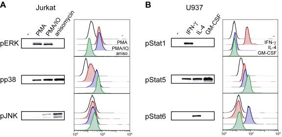
Figure 1 Flow cytometric
analysis of phospho-epitope levels correlates to Western blotting techniques. (A) Jurkat cells were either unstimulated or
treated with PMA (50 nM), PMA (50 nM) and ionomycin (IO, 1 mM), or anisomycin (2 mg/ml) for 10 min at
37°C. Cells were then divided and lysed for Western blot analysis or fixed with
formaldehyde and permeabilized with methanol for flow cytometric analysis
(using optimal techniques, as discussed in the text) with pERK1/2 Alexa (Ax)
488, pp38 Ax647, or pJNK Ax647. Unconjugated mAbs were used for Western blots.
Unstimulated samples appear as open traces in the FACS plots. (B) U937 cells
were left unstimulated or treated with IFN-g (50 ng/ml), IL-4 (10 ng/ml), or GM-CSF (10 ng/ml)
for 10 min at 37°C. The cells were split as above, and analyzed with pStat1
Ax488, pStat5 Ax488, and pStat6 Ax647. Note there is a strong correlation
between Western blotting and flow cytometric analysis with intermediate levels
of pp38 and pStat5 discernible by both methods.