
Anterior Cruciate Ligament (ACL) Reconstruction and Gait
Goals
- To monitor cartilage thinning of both ACL reconstructed and healthy contralateral knees over time using MR imaging and segmentation techniques.
- To study gait changes in ACL reconstructed patients 2 and 4 years post-surgery.
Major Findings
- ACL reconstructed (ACL-R) subjects displayed an external tibial rotational offset throughout stance phase of walking at 2 years post-surgery, which could explain the increased risk for osteoarthritis (OA) in this patient population.
- At two years post operation:
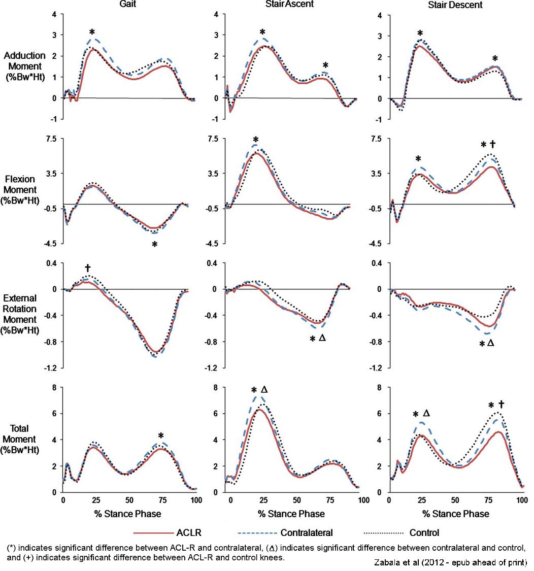
- ACL-R patients have adopted a reduced knee adduction moment when compared to their contralateral, non-injured limb, while walking and ascending and descending stairs.
- In stair ascent and descent, ACL-R patients' contralateral knees have
greater overall total knee joint moments (1st peak) when compared to
matched control (uninjured) subjects.
- Indicates possible compensatory change in ACL-R patients, where the contralateral knee has greater loading maybe due to the lower loading of the ACL-R knee.
- Lower moments in the ACL-R knees also suggest that joint loading might not be the initiating factor for OA in the ACL-R population. The pathway to OA in this population may be initiated by kinematic or biological changes.
- ACL reconstructed knees display lower torsional stiffness in internal tibial rotation.

Representative Publications
- Three-dimensional knee moments of ACL reconstructed and
control subjects during gait, stair ascent, and stair descent.
Zabala ME, Favre J, Scanlan SF, Donahue J, Andriacchi TP. J Biomech. 2012 Nov 7. pii: S0021-9290(12)00608-2. doi: 10.1016/j.jbiomech.2012.10.010. [Epub ahead of print] - Differences in tibial rotation during walking in ACL
reconstructed and healthy contralateral knees.
Scanlan SF, Chaudhari AM, Dyrby CO, Andriacchi TP. J Biomech. 2010 Jun 18;43(9):1817-22. - Zabala M, Scanlan S, Andriacchi T. Alterations in 3D Tibiofemoral Neutral Position Following ACL Deficiency and Reconstruction, the 56th Annual Meeting of the Orthopaedic Research Society, New Orleans, Louisiana, March 6-9, 2010.
- Zabala M, Scanlan S, Donahue J, Andriacchi T. Differences in Active Torsional Stiffness of ACL Reconstructed versus Healthy Contralateral Knees. To be presented at the Orthopaedic Research Society 2012 Meeting in San Francisco, CA.