1:00pm-2:00pm: Presentation and Q&A
2:00pm-2:30pm: Light Refreshments
Talk title: Precision Imaging Guides Cancer Therapy
Historically, many of the gains in medicine have been achieved by uniformly applying medical insights to large groups of patients. A combination of increasing biological understanding of the heterogeneity of disease processes and the concurrent expansion of available selective therapeutic interventions has provided the opportunity to improve group outcomes by optimizing treatment on an individual basis. In many cases, molecular imaging is ideally suited to serve as a biomarker to guide therapy selection and dosing, and to provide an early assessment of efficacy. This presentation highlights by example several areas in which imaging helps determine target engagement, dynamic cellular response to treatment, and the effectiveness of immune modulation in oncologic treatment.

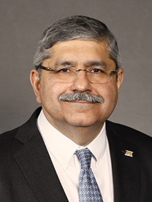
Special PHIND Seminar presented by Dr. Ann Hsing
Title: Stanford WELL for Life Study: A Global Study of Precision Well-being
Speaker: Ann Hsing, PhD
Professor of Medicine
Stanford Prevention Research Center
Stanford Cancer Institute
Department of Health Policy and Research (Epidemiology), by courtesy
Bio: Dr. Ann Hsing is a professor of medicine at Stanford University and co-leader of the population Sciences Program at Stanford Cancer Institute. She is also a professor at Stanford Prevention Research Center and in the Department of Health Research and Policy (Epidemiology, by courtesy). In addition, Dr. Hsing is a faculty fellow for the Center for Innovation in Global Health as well as the Center for Population Health Sciences (PHS) at Stanford Medicine, where she chairs the Pacific Rim Alliance for Population Health, a new multidisciplinary initiative aimed at improving health in the Pacific Rim. Prior to joining Stanford Medicine, Dr. Hsing served as Chief Scientific Officer at the Cancer Prevention Institute of California, a role she assumed after retiring from her post as a tenured intramural investigator at the National Cancer Institute where she served for 23 years. Dr. Hsing received her PhD in epidemiology from the Johns Hopkins University and her master’s degree in biostatistics from the University of California at Los Angeles. At Stanford, she serves as the Principal Investigator of WELL Asia, including longitudinal cohorts in China, Taiwan, and Singapore to investigate socio-behavioral, biochemical, and molecular determinants of well-being. Dr. Hsing has published over 295 peer-reviewed papers and mentored over 65 post-doctoral fellows and junior faculty. In addition to science, Dr. Hsing’s passion is training the next generation of scientists and helping young people succeed in realizing their dreams.
Abstract: As the leader in well-being research, Stanford Prevention Research Center (SPRC) defines well-being as the holistic synthesis of a person’s biological, psychological, and spiritual experiences, resulting from interplay between individuals and their social, economic, and physical environments, that promote living a fulfilling life. Our vision is to improve and sustain health and well-being globally and our mission is to accelerate the science to enhance well-being. To accomplish this, we established the Stanford WELL for Life Study, an international study that uses novel methods to define, assess, and promote the multiple dimensions of well-being in the U.S. and globally. The Stanford WELL for Life Study uses a data-driven approach to define and measure well-being, identify factors related to well-being, and evaluate the impact of interventions on well-being. Currently, there are five study sites—the San Francisco Bay Area, China (Hangzhou), Taiwan (Taipei), Singapore, and Thailand (Bangkok)—with more than 24,000 individuals enrolled to date. We have collected data on 400-1,000 variables per individual and obtained biospecimens from 80% of participants for future molecular investigations. To assess well-being, SPRC developed a de novo multi-dimension survey (the Stanford WELL for Life Scale) that measures ten domains of well-being and a total well-being score. These ten domains, which emerged from our unique and extensive qualitative data, include: social connectedness, lifestyle and daily practices, stress and resilience, experience of emotions, physical health, purpose and meaning, sense of self, financial security and satisfaction, spirituality and religiosity, exploration and creativity. At the PHIND seminar, I will share with you the genesis and evolution of the Stanford WELL for Life Study and our exciting preliminary data.

PHIND Seminar August
Mehmet Ozgun
“Extracellular Vesicles for Broad Applications in Medicine and Cancer”
About Mehmet O. Ozen, PhD
Dr. Ozen is a Postdoctoral Research Fellow at Canary Center for Cancer Early Detection / Radiology Department at Stanford University. He works with Prof. Utkan Demirci on simple solutions for complex problems in medicine, combining microfluidics and bioengineering principles. He received his BS and PhD in Bioengineering from Ege University.
Abstract
Extracellular vesicles (EVs) are lipid bi-layered nanoparticles shed from the cells that carry RNA, DNA, transmembrane and cytosolic proteins. The variety in EV size, cargo and origin attracted researchers to decipher the mechanisms that have been involved in packaging, secretion, uptake and roles of EVs on cells in vivo and in vitro, lightening the path for biomarker studies for diagnosis, prognosis, therapy and therapy monitoring. They are one of the many means that cells use to communicate with neighboring and distant cells and tissues. With improvements in next-generation sequencing technologies and increased resolution of mass spectrometry for proteomic analysis, EVs have been shown to take role in angiogenesis, epithelial-to-mesenchymal transition, stemness in cancer, malignancy, metastasis and drug resistance.
Although exosomes show unprecedented promising advantages over other biotargets in the circulation for clinical use, a major challenge rapidly emerging in the field of EV utilization for clinical and non-clinical applications is the absence of reproducible, inexpensive and robust tools for efficient sorting and isolation of EV populations at a high yield. The field lacks a clear consensus over an optimum approach or a tool for isolation of EVs avoiding contamination with many other proteins and such other biostructures and reproducible procedures for downstream analysis of EV cargo and content. Existing approaches for EV isolation include a variety of methods. Additionally, methods for the exosome-derived analyte isolation, library preparation for sequencing, and downstream analysis including genomic, proteomic and metabolic analysis are highly varied. Hence, there is a need for well-developed experimental tools, interlaboratory evaluations and in-depth descriptions of experimental steps and designs to ensure reliable, robust and reproducible experiments and tools.
In this talk, we will describe a new technique, i.e., Exosome Total Isolation Chip (ExoTIC), that is developed in our lab to isolate EVs and EV subpopulations from a variety of sample types including plasma and culture media. We will present further downstream genomic and proteomic analysis of these EVs focusing on applications in cancer and cardiovascular disorders.


PHIND Seminar Series October: ‘Progression of Clonal Hematopoiesis of Indeterminate Potential to Acute Myeloid Leukemia’
Ravi Majeti, MD, Ph.D.
Professor of Medicine
Chief, Division of Hematology
Institute for Stem Cell Biology and Regenerative Medicine
Stanford University
Munzer Auditorium (B060), Beckman Center
11:00am-12:00pm – Seminar and Discussion
12:00pm-12:15pm – Reception (light refreshments provided)
RSVP Here: https://www.onlineregistrationcenter.com/register/222/page1.asp?m=298&c=39
ABSTRACT: Myeloid malignancies are cancers of the blood lineage including myeloproliferative neoplasms (MPN), myelodysplastic syndromes (MDS), and acute myeloid leukemia (AML) with more than 40,000 new diagnoses annually in the United States. These diseases cause significant morbidity and mortality due to associated bone marrow failure leading to anemia, bleeding, and infections, and are currently treated with targeted therapies, chemotherapy, and allogeneic bone marrow transplantation. Next generation DNA sequencing has determined the spectrum of mutations associated with these cancers and has found that most cases are associated with multiple mutations that cooperate to cause disease. In our prior studies, we determined that these mutations are serially acquired in clones of self-renewing pre-cancerous/pre-leukemic blood stem cells. Separate studies analyzed blood sequencing data from large cohorts of individuals without disease and found these pre-leukemic mutations occur in the general population with increasing frequency and incidence with age. As only a minor subset of these individuals eventually progressed to develop myeloid malignancy, this entity was termed clonal hematopoiesis of indeterminate potential (CHIP). One major issue with implications for the transition from health to disease is to understand what factors influence the progression from CHIP to myeloid malignancy. In order to investigate this question, we have developed models for CHIP/pre-leukemia through the CRISPR-mediated engineering of normal human blood stem and progenitor cells. By introducing mutations in the TET2 and ASXL1 genes that are commonly mutated in CHIP, we have established models for the cell intrinsic processes of progression to myeloid malignancy and are now poised to examine cell extrinsic processes that can affect such progression. Establishing these models is key to investigating measures to eventually prevent development of myeloid malignancy.

PHIND Seminar Series November: ‘ What You Always Wanted to Know about Economics, Payer Coverage, and Big Data for Precision Health – But Were Afraid to Ask’
Kathryn Phillips, Ph.D.
Professor of Health Economics
Founding Director of the UCSF Center for Translational and Policy Research on Personalized Medicine (TRANSPERS)
Department of Clinical Pharmacy
UCSF
Li Ka Shing Center, LK101
11:00am-12:00pm – Seminar and Discussion
12:00pm-12:15pm – Reception (light refreshments provided)
RSVP Here: https://www.onlineregistrationcenter.com/KathrynPhillips
ABSTRACT: Precision Health offers an opportunity to achieve “high value care” through innovative approaches. However, in order to fulfill this objective, we must demonstrate its economic value, someone must be willing to pay the costs, and there has to be data available to provide the needed evidence. In this talk, I will draw on my research over the past decade examining (1) how to measure the value of complex technologies such as Precision Health, (2) what payers cover and how they decide to provide coverage, and (3) how Big Data can be leveraged. I will also describe “lessons learned” about successful adoption from working with dozens of start-ups, VCs, and biotech companies. The talk will illustrate these issues using the case study of “liquid biopsy” – a potentially transformative technology that illustrates both the opportunities and challenges for Precision Health.

Integrative Biomedical Imaging Informatics at Stanford (IBIIS) and Center for Artificial Intelligence in Medicine & Imaging (AIMI) Seminar: “AI-Aided Diagnostic and Prognostic Tools for Prostate Cancer”
Okyaz Eminaga, MD, PhD
Postdoctoral Research Fellow, Urology
Biomedical Data Sciences
Stanford University
James H. Clark Center, S360
12:00pm-1:00pm – Seminar and Discussion (light refreshments provided)
Join via Zoom: https://stanford.zoom.us/j/613898274
ABSTRACT: Prostate Cancer exhibits different clinical behavior, ranging from indolent to lethal disease. A critical clinical need is identifying characteristics that distinguish indolent from advanced disease to direct treatment to the latter. The recent renaissance of artificial intelligence (AI) research uncovered the potential of AI to improve clinical decision making. In this seminar, we will go through the potential of AI to enhance the diagnosis and the prognosis of prostate cancer using magnetic resonance images, clinical data, and histology images. We will stress the challenges and benefits of having such AI-based solutions in clinical routine.
ABOUT: Dr. Eminaga passed his medical examination (Staatsexamen) 2009 and received his Ph.D. in Medicine 2010 from University of Muenster (major topic: medical informatics) under the supervision of Professor Dr. Axel Semjonow (one of the pioneer physician-scientists and biomarker researcher who worked on the standardization of PSA measurement for prostate cancer which is used nowadays) and Professor Dr. Martin Dugas (who is the head of European Research Center for Information Systems and one of the most influential professors in medical informatics in Europe). For those who don’t know the institute of medical informatics in Muenster. The systematized Nomenclature of Human and Veterinary Medicine (SNOMED), which is now used worldwide in medical information systems, was initiated by this institute more than 30 years ago.
His doctoral dissertation presented a novel documentation architecture for clinical data and imaging called cMDX (clinical map document) that facilitates the concept of the single-source information system for clinical data storage and analysis, and is successfully used in clinical routine for generating the pathology reports with graphical information about the spatial tumor extent for prostatectomy specimens since 2009 at the prostate center of University Hospital Muenster. This work has been also utilized for more than 20 studies related to genomics, translational medicine, epidemiology, urology, radiology, and pathology. Dr. Eminaga also established the biobanking information management system to manage the samples of one of the largest biobanks for prostate cancer in Europe. This biobank is also part of the European P-Mark network for prostate cancer-related biorepositories initiated by Oxford University.
Dr. Eminaga completed his residency in Urology in the University Hospital of Cologne (Germany) with a major focus on uro-oncology. He was also a research fellow in Prostate Center of University Hospital Muenster, doing research in biomarkers, biobanking infrastructure, epidemiology and histopathology. During his residency fellowship, he further evaluated the role of certain miRNA in prostate cancer development under the supervision of the molecular biologist Dr. Warnecke-Eberz. After his residency, he started a research scholarship at the laboratory of Dr. Brooks, doing genomic research and bioinformatics for research topics related to prostate cancer evolution. Now, his current interests have expanded to statistical learning, medical imaging informatics, and integrative data analysis.
He is the recipient of 3 highly-competitive scholarships and his works have been recognized at national and international levels e.g., by the European Association of Urology. Currently, he is an early-investigator research awardee for prostate cancer managed by the department of defense and works on developing decision-aided tools for diagnosis and prognosis of prostate cancer.
Follow us on Twitter: @StanfordIBIIS and @StanfordAIMI
http://ibiis.stanford.edu/events/seminars/2019seminars.html

“Messaging in the Age of Microtargeting”
John Stafford
Assistant Vice President
Digital Strategy
Stanford University
Bjorn Carey
Senior Director
Digital Strategy
Stanford University
Join via Zoom: https://stanford.zoom.us/j/400566542
Abstract:
Communications has become increasingly data-driven, targeted, and personalized. This has changed how Stanford analyzes communications opportunities from a research perspective and how it engages with relevant audiences. In this presentation, John and Bjorn will share the data and communications strategy underlying three communications initiatives and the resulting execution. They will also provide practical advice for individual thought leadership and communications in this dynamic environment.
About:
John Stafford, MA ’06, is currently Assistant Vice President for Digital Strategy at Stanford, the most senior digital communications role in the university. John is responsible for all aspects of creating a world-class digital communications function: setting the group’s strategy, building analytics and insight programs, counseling on crisis communications, leading multi-channel messaging initiatives, and advising colleagues across the University. He received a Master’s Degree in Communication from Stanford, a B.A. in History from the University of San Francisco, and was a founding advisor to Stanford Medicine X.
Refreshments will be provided.

“Algorithm Development Lifecycle in Medical Imaging:
Current State and Considerations for the Future”
Luciano M. Prevedello, MD, MPH
Vice-Chair for Medical Informatics and Augmented Intelligence in Imaging
Division Chief, Medical Imaging Informatics
Director, 3D and Advanced Visualization Lab
Associate Professor, Division of Neuroradiology,
Department of Radiology
Ohio State University Wexner Medical Center
Join via Zoom: https://stanford.zoom.us/j/267814863
Abstract:
This presentation will describe some of the most important considerations involved in creating algorithms in medical imaging from inception to deployment as well as continued model improvement and/or monitoring. Examples of experience to date from the OSU laboratory for augmented intelligence in imaging will be provided. New paradigms in model creation and the role of image challenge competitions will also be covered. Current issues with model validation and generalizability will also be introduced as well as considerations for future work in this area.
Refreshments will be provided.

“A Deep Learning Framework for Efficient Registration of MRI and Histopathology Images of the Prostate”
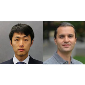
Wei Shao, PhD
Postdoctoral Research Fellow
Department of Radiology
Stanford University
“Applications of Generative Adversarial Networks (GANs) in Medical Imaging”
Saeed Seyyedi, PhD
Paustenbach Research Fellow
Department of Radiology
Stanford University
Join via Zoom: https://stanford.zoom.us/j/593016899
Refreshments will be provided
ABSTRACT (Shao)
Magnetic resonance imaging (MRI) is an increasingly important tool for the diagnosis and treatment of prostate cancer. However, MRI interpretation suffers from high interobserver variability and often misses clinically significant cancers. Registration of histopathology images from patients who have undergone surgical resection of the prostate onto pre-operative MRI images allows direct mapping of cancer location onto MR images. This is essential for the discovery and validation of novel prostate cancer signatures on MRI. Traditional registration approaches can be computationally expensive and require a careful choice of registration hyperparameters. We present a deep learning-based pipeline to accelerate and simplify MRI-histopathology image registration in prostate cancer. Our pipeline consists of preprocessing, transform estimation by deep neural networks, and postprocessing. We refined the registration neural networks, originally trained with 19,642 natural images, by adding 17,821 medical images of the prostate to the training set. The pipeline was evaluated using 99 prostate cancer patients. The addition of the images to the training set significantly (p < 0.001) improved the Dice coefficient and reduced the Hausdorff distance. Our pipeline also achieved comparable accuracy to an existing state-of-the-art algorithm while reducing the computation time from 4.4 minutes to less than 2 seconds.
ABSTRACT (Seyyedi)
Generative adversarial networks (GANs) are advanced types of neural networks where two networks are trained simultaneously to perform two tasks of generation and discrimination. GANs have gained a lot of attention to tackle well known and challenging problems in computer vision applications including medical image analysis tasks such as medical image de-noising, detection and classification, segmentation and reconstruction.In this talk, we will introduce some of the recent advancements of GANs in medical imaging applications and will discuss the recent developments of GAN models to resolve real world imaging challenges.

MIPS Seminar: “Tiny Bubbles, Big Impact: Exploring applications of nanobubbles in ultrasound molecular imaging and therapy”
Agata A. Exner, Ph.D.
Professor of Radiology and Biomedical Engineering
Department of Radiology
Case Western Reserve
Location: Beckman Center, B230
2:00pm – 3:00pm Seminar & Discussion
ABSTRACT
Sub-micron shell stabilized gas bubbles (aka nanobubbles (NB) or ultrafine bubbles) have gained momentum as a robust contrast agent for molecular imaging and therapy using ultrasound. The small size, extended stability and high concentration of nanobubbles make them an ideal tool for new applications of contrast enhanced ultrasound and ultra-
sound-mediated therapy, especially in oncology-related problems. Compared to microbub-bles, nanobubbles can provide superior tumor delineation, identify biomarkers on the vascu-lature and on tumors cells and facilitate drug and gene delivery into tumor tissue. The pat-terns of tissue enhancement under nonlinear ultrasound imaging of nanobubbles are distinct from conventional microbubbles especially in tissues exhibiting vascular hyperper-meability. Specifically, NB kinetics, quantified via time intensity curve analysis, typically show a marked delay in the washout rate and significantly increased area under the curve compared to larger bubbles. This effect is further enhanced by molecular targeting to cellular biomarkers, such as the prostate specific membrane antigen (PSMA) or the receptor protein tyrosine phosphatase, PTPmu. The unique contrast enhancement dynamics of nanobubbles are likely to be a result of direct bubble extravasation and prolonged retention of intact bubbles in target tissue. Thus, understanding the underlying mechanisms behind the unique nanobubble behavior can be the driver of significant future innovations in contrast enhanced ultrasound imaging applications. This presentation will discuss the fundamental challenges with nanobubble formulation and characterization and will showcase how the unique fea-tures of nanobubbles can be leveraged to improve disease detection and treatment using ultrasound.