Abstract: Gene editing with CRISPR technology is transforming biology. Understanding the underlying chemical mechanisms of RNA-guided DNA and RNA cleavage provides a foundation for both conceptual advances and technology development. I will discuss how bacterial CRISPR adaptive immune systems inspire creation of powerful genome editing tools, enabling advances in both fundamental biology and applications in medicine. I will also discuss the ethical challenges of some of these applications with a focus on what our decisions now might mean for future generations.
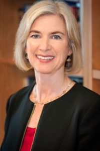
About: MIPS IMAGinING THE FUTURE seminar series is aimed at catalyzing interdisciplinary discussions in all area of medicine and disease. The seminar series is open and free to everyone in the Stanford community, as well as anyone from the surrounding community, companies or institutions. Our next seminar will host Dr. Jennifer Doudna, Professor of Chemistry, Biochemistry & Molecular Biology, &Li Ka Shing Chancellor’s Professor in Biomedical and Health, University of California, Berkeley; for her presentation on the “World of CRISPR: Editing Genomes and Altering Our Future”.
More Information: http://med.stanford.edu/radiology/imagining-the-future.html
Register: https://www.onlineregistrationcenter.com/JenniferDoudna

PHIND Seminar Series October: ‘Progression of Clonal Hematopoiesis of Indeterminate Potential to Acute Myeloid Leukemia’
Ravi Majeti, MD, Ph.D.
Professor of Medicine
Chief, Division of Hematology
Institute for Stem Cell Biology and Regenerative Medicine
Stanford University
Munzer Auditorium (B060), Beckman Center
11:00am-12:00pm – Seminar and Discussion
12:00pm-12:15pm – Reception (light refreshments provided)
RSVP Here: https://www.onlineregistrationcenter.com/register/222/page1.asp?m=298&c=39
ABSTRACT: Myeloid malignancies are cancers of the blood lineage including myeloproliferative neoplasms (MPN), myelodysplastic syndromes (MDS), and acute myeloid leukemia (AML) with more than 40,000 new diagnoses annually in the United States. These diseases cause significant morbidity and mortality due to associated bone marrow failure leading to anemia, bleeding, and infections, and are currently treated with targeted therapies, chemotherapy, and allogeneic bone marrow transplantation. Next generation DNA sequencing has determined the spectrum of mutations associated with these cancers and has found that most cases are associated with multiple mutations that cooperate to cause disease. In our prior studies, we determined that these mutations are serially acquired in clones of self-renewing pre-cancerous/pre-leukemic blood stem cells. Separate studies analyzed blood sequencing data from large cohorts of individuals without disease and found these pre-leukemic mutations occur in the general population with increasing frequency and incidence with age. As only a minor subset of these individuals eventually progressed to develop myeloid malignancy, this entity was termed clonal hematopoiesis of indeterminate potential (CHIP). One major issue with implications for the transition from health to disease is to understand what factors influence the progression from CHIP to myeloid malignancy. In order to investigate this question, we have developed models for CHIP/pre-leukemia through the CRISPR-mediated engineering of normal human blood stem and progenitor cells. By introducing mutations in the TET2 and ASXL1 genes that are commonly mutated in CHIP, we have established models for the cell intrinsic processes of progression to myeloid malignancy and are now poised to examine cell extrinsic processes that can affect such progression. Establishing these models is key to investigating measures to eventually prevent development of myeloid malignancy.
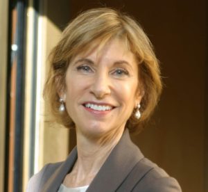
PHIND Seminar Series November: ‘ What You Always Wanted to Know about Economics, Payer Coverage, and Big Data for Precision Health – But Were Afraid to Ask’
Kathryn Phillips, Ph.D.
Professor of Health Economics
Founding Director of the UCSF Center for Translational and Policy Research on Personalized Medicine (TRANSPERS)
Department of Clinical Pharmacy
UCSF
Li Ka Shing Center, LK101
11:00am-12:00pm – Seminar and Discussion
12:00pm-12:15pm – Reception (light refreshments provided)
RSVP Here: https://www.onlineregistrationcenter.com/KathrynPhillips
ABSTRACT: Precision Health offers an opportunity to achieve “high value care” through innovative approaches. However, in order to fulfill this objective, we must demonstrate its economic value, someone must be willing to pay the costs, and there has to be data available to provide the needed evidence. In this talk, I will draw on my research over the past decade examining (1) how to measure the value of complex technologies such as Precision Health, (2) what payers cover and how they decide to provide coverage, and (3) how Big Data can be leveraged. I will also describe “lessons learned” about successful adoption from working with dozens of start-ups, VCs, and biotech companies. The talk will illustrate these issues using the case study of “liquid biopsy” – a potentially transformative technology that illustrates both the opportunities and challenges for Precision Health.


“Messaging in the Age of Microtargeting”
John Stafford
Assistant Vice President
Digital Strategy
Stanford University
Bjorn Carey
Senior Director
Digital Strategy
Stanford University
Join via Zoom: https://stanford.zoom.us/j/400566542
Abstract:
Communications has become increasingly data-driven, targeted, and personalized. This has changed how Stanford analyzes communications opportunities from a research perspective and how it engages with relevant audiences. In this presentation, John and Bjorn will share the data and communications strategy underlying three communications initiatives and the resulting execution. They will also provide practical advice for individual thought leadership and communications in this dynamic environment.
About:
John Stafford, MA ’06, is currently Assistant Vice President for Digital Strategy at Stanford, the most senior digital communications role in the university. John is responsible for all aspects of creating a world-class digital communications function: setting the group’s strategy, building analytics and insight programs, counseling on crisis communications, leading multi-channel messaging initiatives, and advising colleagues across the University. He received a Master’s Degree in Communication from Stanford, a B.A. in History from the University of San Francisco, and was a founding advisor to Stanford Medicine X.
Refreshments will be provided.

MIPS Seminar: “Tiny Bubbles, Big Impact: Exploring applications of nanobubbles in ultrasound molecular imaging and therapy”
Agata A. Exner, Ph.D.
Professor of Radiology and Biomedical Engineering
Department of Radiology
Case Western Reserve
Location: Beckman Center, B230
2:00pm – 3:00pm Seminar & Discussion
ABSTRACT
Sub-micron shell stabilized gas bubbles (aka nanobubbles (NB) or ultrafine bubbles) have gained momentum as a robust contrast agent for molecular imaging and therapy using ultrasound. The small size, extended stability and high concentration of nanobubbles make them an ideal tool for new applications of contrast enhanced ultrasound and ultra-
sound-mediated therapy, especially in oncology-related problems. Compared to microbub-bles, nanobubbles can provide superior tumor delineation, identify biomarkers on the vascu-lature and on tumors cells and facilitate drug and gene delivery into tumor tissue. The pat-terns of tissue enhancement under nonlinear ultrasound imaging of nanobubbles are distinct from conventional microbubbles especially in tissues exhibiting vascular hyperper-meability. Specifically, NB kinetics, quantified via time intensity curve analysis, typically show a marked delay in the washout rate and significantly increased area under the curve compared to larger bubbles. This effect is further enhanced by molecular targeting to cellular biomarkers, such as the prostate specific membrane antigen (PSMA) or the receptor protein tyrosine phosphatase, PTPmu. The unique contrast enhancement dynamics of nanobubbles are likely to be a result of direct bubble extravasation and prolonged retention of intact bubbles in target tissue. Thus, understanding the underlying mechanisms behind the unique nanobubble behavior can be the driver of significant future innovations in contrast enhanced ultrasound imaging applications. This presentation will discuss the fundamental challenges with nanobubble formulation and characterization and will showcase how the unique fea-tures of nanobubbles can be leveraged to improve disease detection and treatment using ultrasound.

MIPS Seminar
2:00-2:45 PM | Prof. Pawel Moskal
“Positronium Imaging with the J-PET Scanner”
Head of the Department of Experimental Particle Physics and Applications
Marian Smoluchowski Institute of Physics
Jagiellonian University, 30-348 Krakow, Poland
2:45-3:30 PM | Prof. Ewa Stepien
“Preclinical studies of positronium and extracellular vesicles biomarkers”
Head of the Department of Medical Physics
Marian Smoluchowski Institute of Physics
Jagiellonian University, 30-348 Krakow, Poland
ABSTRACT
As modern medicine develops towards personalized treatment of patients, there is a need for highly specific and sensitive tests to diagnose disease. Our research aims at improvement of specificity of positron emission tomography (PET) in assessment of cancer by use of positronium as a theranostic agent. During PET scanning about 40% of positron annihilations occur through the creation of positronium. “Positronium,” which may be formed in human tissues in the intramolecular spaces, is an exotic atom composed of an electron from tissue and the positron emitted by the radioinuclide. Positronium decay in the patient body is sensitive to the nanostructure and metabolism of human tissues. This phenomenon is not used in present PET diagnostics, yet it is in principle possible to exploit such environment modified properties of positronium as diagnostic biomarkers for cancer assessment. Our first in-vitro studies have shown differences of the positronium mean lifetime and production probability in healthy and cancerous tissues, indicating that they may be used as indicators for in-vivo cancer classification. For the application in medical diagnostics, the properties of positronium atoms need to be determined in a spatially resolved manner. For that purpose we have developed a method of positronium lifetime imaging in which the lifetime and position of positronium atoms are determined on an event-by-event basis. This method requires application of β+ decaying isotope that also emits a prompt gamma ray. We will argue that with total-body PET scanners, the sensitivity of positronium lifetime imaging, which requires coincident registration of the back-to-back annihilation photons and the prompt gamma, is comparable to the sensitivities for metabolic imaging with standard PET scanners.
Our research involves also development of diagnostic methods based on the extracellular vesicles (EVs), which are micro and nano-sized, closed membrane fragments. They are produced by native cells to facilitate the transfer of different signaling factors, structural proteins, nucleic acids or lipids even to distant cells. They are present in all body fluids and they are specific to their parental cells.
Our presentation will be divided into two parts. In the first, the method of positronium imaging and the pilot positronium images obtained with the J-PET detector (the first PET system built based on plastic scintillators) will be reported. This part of the presentation will include also description and perspectives of development of the J-PET technology in view of total-body PET imaging. The second part will concern preliminary results of the preclinical studies of positronium properties in cancerous and healthy tissues sampled from patients as well as in the frozen and living healthy and cancer skin cells in-vitro. The second part will include also description of the novel method for the diagnosis of diabetes and melanoma based on EVs used as biomarkers and drug delivery systems.
References:
P. Moskal, …. E. Ł. Stępień et al., Phys. Med. Biol. 64 (2019) 055017
- Moskal, B. Jasinska, E. Ł. Stępień, S. Bass, Nature Reviews Physics 1 (2019) 527
- Roman M… .E. Ł. Stępień, Nanomedicine 17 (2019) 137
- Ł. Stępień et al., Theranostics 8 (2018) 3874
Hosted by: Craig Levin, Ph.D.
Sponsored by the Molecular Imaging Program at Stanford and the Department of Radiology
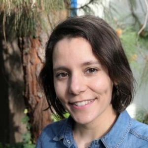
PHIND Seminar Series: “Prediction of Future Lymphoma Development Based on DNA Methylation Profiles from Peripheral Blood”
Almudena Espin Perez, PhD
Postdoctoral Research Fellow
Biomedical Informatics
Stanford University
Beckman Center, Munzer Auditorium (B060)
12:00pm – 1:00pm Seminar & Discussion
1:00pm – 1:15pm Reception & Light Refreshments
RSVP here: https://www.onlineregistrationcenter.com/APerez
ABSTRACT
Subjects with Non-Hodgkin Lymphoma (NHL) have abnormal lymphocytes that multiply and accumulate to form tumors in the lymph nodes and other organs. Currently, there are no predictive models with high performance that can predict the risk of developing NHL.
We present a computational framework that accurately predicts future (up to 16 years) NHL from a signature based on DNA methylation profiles of peripheral blood samples. We studied differences in specific DNA methylation levels from blood samples between future NHL group and the control group (470 samples) from two prospective cohorts. We developed a predictive model using advanced artificial intelligence methods for NHL diagnosis based on a set of key CpG sites. The validation tests showed that our signature 1) predicts mainly “control” in an independent population of 656 healthy subjects, 2) predicts “future case” with extremely accurate performance in tissue samples from four independent NHL cohorts (662, 29, 31 and 29 subjects), with one of the cohorts (662 subjects) corresponding to children with B-cell lymphoma, 3) predicts mostly healthy in a cohort of children with 74 children in remission, 4) works for both HIV positive subjects and HIV negative subjects, 5) yields almost perfect predictions regardless of the NHL subtype, and 6) is 84% accurate at predicting T-cell lymphoma in children, despite its derivation in B-cell lymphoma in adults.
ABOUT
Almudena Espin Perez’s interests include developing algorithms and novel computational methods for early cancer detection. High-throughput technologies in the field of molecular biology are generating huge amounts of biological data and transforming the scientific landscape. A major focus of her research is on building computational methods to 1) study genomics and epigenetic data 2) integrate genomics and imaging data at single-cell level resolution and 3) leverage existing large-scale transcriptomic datasets to address relevant biological questions by developing computational deconvolution tools to infer the abundance of different cell types from mixed cell populations. Dr. Perez aims to improve the understanding of the molecular mechanisms behind cancer development, which could potentially lead to biomarker discovery and improve early detection, treatment strategies and decision-making.
Hosted by: Sanjiv Sam Gambhir, M.D., Ph.D.
Sponsored by the PHIND Center and the Department of Radiology
Please note this seminar is now cancelled and will be rescheduled for a later date.
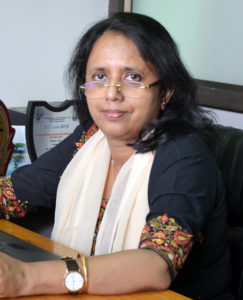
MIPS Seminar: Investigating and Imaging key molecular switches associated with Acquirement of Platinum-Taxol resistance in Epithelial Ovarian Cancer
Pritha Ray, Ph.D.
Principal Investigator & Scientific Officer F
Imaging Cell Signaling & Therapeutics Lab
ACTREC, Tata Memorial Center
Navi Mumbai, India

Please note this seminar is now cancelled and will be rescheduled for a future date. Please contact Ashley Williams (ashleylw@stanford.edu) with any questions or concerns. Thank you for your understanding!
PHIND Seminar Series: “A Stroke Monitoring and Alert System for a Future Without Late Presentation”
Orestis Vardoulis, Ph.D.
Postdoctoral Research Fellow
Pediatric Surgery
Stanford University