Location & Timing
August 5, 2020
8:30am-4:30pm
Livestream: details to come
This event is free and open to all!
Registration and Event details
Overview
Advancements of machine learning and artificial intelligence into all areas of medicine are now a reality and they hold the potential to transform healthcare and open up a world of incredible promise for everyone. Sponsored by the Stanford Center for Artificial Intelligence in Medicine and Imaging, the 2020 AIMI Symposium is a virtual conference convening experts from Stanford and beyond to advance the field of AI in medicine and imaging. This conference will cover everything from a survey of the latest machine learning approaches, many use cases in depth, unique metrics to healthcare, important challenges and pitfalls, and best practices for designing building and evaluating machine learning in healthcare applications.
Our goal is to make the best science accessible to a broad audience of academic, clinical, and industry attendees. Through the AIMI Symposium we hope to address gaps and barriers in the field and catalyze more evidence-based solutions to improve health for all.

Judy Gichoya, MD
Assistant Professor
Emory University School of Medicine
Measuring Learning Gains in Man-Machine Assemblage When Augmenting Radiology Work with Artificial Intelligence
Abstract
The work setting of the future presents an opportunity for human-technology partnerships, where a harmonious connection between human-technology produces unprecedented productivity gains. A conundrum at this human-technology frontier remains – will humans be augmented by technology or will technology be augmented by humans? We present our work on overcoming the conundrum of human and machine as separate entities and instead, treats them as an assemblage. As groundwork for the harmonious human-technology connection, this assemblage needs to learn to fit synergistically. This learning is called assemblage learning and it will be important for Artificial Intelligence (AI) applications in health care, where diagnostic and treatment decisions augmented by AI will have a direct and significant impact on patient care and outcomes. We describe how learning can be shared between assemblages, such that collective swarms of connected assemblages can be created. Our work is to demonstrate a symbiotic learning assemblage, such that envisioned productivity gains from AI can be achieved without loss of human jobs.
Specifically, we are evaluating the following research questions: Q1: How to develop assemblages, such that human-technology partnerships produce a “good fit” for visually based cognition-oriented tasks in radiology? Q2: What level of training should pre-exist in the individual human (radiologist) and independent machine learning model for human-technology partnerships to thrive? Q3: Which aspects and to what extent does an assemblage learning approach lead to reduced errors, improved accuracy, faster turn-around times, reduced fatigue, improved self-efficacy, and resilience?
Zoom: https://stanford.zoom.us/j/93580829522?pwd=ZVAxTCtEdkEzMWxjSEQwdlp0eThlUT09

Ge Wang, PhD
Clark & Crossan Endowed Chair Professor
Director of the Biomedical Imaging Center
Rensselaer Polytechnic Institute
Troy, New York
Abstract:
AI-based tomography is an important application and a new frontier of machine learning. AI, especially deep learning, has been widely used in computer vision and image analysis, which deal with existing images, improve them, and produce features. Since 2016, deep learning techniques are actively researched for tomography in the context of medicine. Tomographic reconstruction produces images of multi-dimensional structures from externally measured “encoded” data in the form of various transforms (integrals, harmonics, and so on). In this presentation, we provide a general background, highlight representative results, and discuss key issues that need to be addressed in this emerging field.
About:
AI-based X-ray Imaging System (AXIS) lab is led by Dr. Ge Wang, affiliated with the Department of Biomedical Engineering at Rensselaer Polytechnic Institute and the Center for Biotechnology and Interdisciplinary Studies in the Biomedical Imaging Center. AXIS lab focuses on innovation and translation of x-ray computed tomography, optical molecular tomography, multi-scale and multi-modality imaging, and AI/machine learning for image reconstruction and analysis, and has been continuously well funded by federal agencies and leading companies. AXIS group collaborates with Stanford, Harvard, Cornell, MSK, UTSW, Yale, GE, Hologic, and others, to develop theories, methods, software, systems, applications, and workflows.

Targeted violence continues against Black Americans, Asian Americans, and all people of color. The department of radiology diversity committee is running a racial equity challenge to raise awareness of systemic racism, implicit bias and related issues. Participants will be provided a list of resources on these topics such as articles, podcasts, videos, etc., from which they can choose, with the “challenge” of engaging with one to three media sources prior to our session (some videos are as short as a few minutes). Participants will meet in small-group breakout sessions to discuss what they’ve learned and share ideas.
Please reach out to Marta Flory, flory@stanford.edu with questions. For details about the session, including recommended resources and the Zoom link, please reach out to Meke Faaoso at mfaaoso@stanford.edu.

Radiology Department-Wide Research Meeting
• Research Announcements
• Mirabela Rusu, PhD – Learning MRI Signatures of Aggressive Prostate Cancer: Bridging the Gap between Digital Pathologists and Digital Radiologists
• Akshay Chaudhari, PhD – Data-Efficient Machine Learning for Medical Imaging
Location: Zoom – Details can be found here: https://radresearch.stanford.edu
Meetings will be the 3rd Friday of each month.
Hosted by: Kawin Setsompop, PhD
Sponsored by: the the Department of Radiology

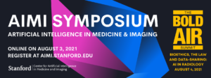
Stanford AIMI Director Curt Langlotz and Co-Directors Matt Lungren and Nigam Shah invite you to join us on August 3 for the 2021 Stanford Center for Artificial Intelligence in Medicine and Imaging (AIMI) Symposium. The virtual symposium will focus on the latest, best research on the role of AI in diagnostic excellence across medicine, current areas of impact, fairness and societal impact, and translation and clinical implementation. The program includes talks, interactive panel discussions, and breakout sessions. Registration is free and open to all.
Also, the 2nd Annual BiOethics, the Law, and Data-sharing: AI in Radiology (BOLD-AIR) Summit will be held on August 4, in conjunction with the AIMI Symposium. The summit will convene a broad range of speakers in bioethics, law, regulation, industry groups, and patient safety and data privacy, to address the latest ethical, regulatory, and legal challenges regarding AI in radiology.

Regina Barzilay, PhD
School of Engineering Distinguished Professor for AI and Health
Electrical Engineering and Computer Science Department
AI Faculty Lead at Jameel Clinic for Machine Learning in Health
Computer Science and Artificial Intelligence Lab
Massachusetts Institute of Technology
Abstract:
In this talk, I will present methods for future cancer risk from medical images. The discussion will explore alternative ways to formulate the risk assessment task and focus on algorithmic issues in developing such models. I will also discuss our experience in translating these algorithms into clinical practice in hospitals around the world.
Keynote:
Self-Supervision for Learning from the Bottom Up
Why do self-supervised learning? A common answer is: “because data labeling is expensive.” In this talk, I will argue that there are other, perhaps more fundamental reasons for working on self-supervision. First, it should allow us to get away from the tyranny of top-down semantic categorization and force meaningful associations to emerge naturally from the raw sensor data in a bottom-up fashion. Second, it should allow us to ditch fixed datasets and enable continuous, online learning, which is a much more natural setting for real-world agents. Third, and most intriguingly, there is hope that it might be possible to force a self-supervised task curriculum to emerge from first principles, even in the absence of a pre-defined downstream task or goal, similar to evolution. In this talk, I will touch upon these themes to argue that, far from running its course, research in self-supervised learning is only just beginning.

Saeed Hassanpour, PhD
Associate Professor of Biomedical Data Science
Associate Professor of Epidemiology
Associate Professor of Computer Science
Dartmouth Geisel School of Medicine
Deep Learning for Histology Images Analysis
Abstract:
With the recent expansions of whole-slide digital scanning, archiving, and high-throughput tissue banks, the field of digital pathology is primed to benefit significantly from deep learning technology. This talk will cover several applications of deep learning for characterizing histopathological patterns on high-resolution microscopy images for cancerous and precancerous lesions. Furthermore, the current challenges for building deep learning models for pathology image analysis will be discussed and new methodological advances to address these bottlenecks will be presented.
About:
Dr. Saeed Hassanpour is an Associate Professor in the Departments of Biomedical Data Science, Computer Science, and Epidemiology at Dartmouth College. His research is focused on machine learning and multimodal data analysis for precision health. Dr. Hassanpour has led multiple NIH-funded research projects, which resulted in novel machine learning and deep learning models for medical image analysis and clinical text mining to improve diagnosis, prognosis, and personalized therapies. Before joining Dartmouth, he worked as a Research Engineer at Microsoft. Dr. Hassanpour received his Ph.D. in Electrical Engineering with a minor in Biomedical Informatics from Stanford University and completed his postdoctoral training at Stanford Center for Artificial Intelligence in Medicine & Imaging.

Indrani Bhattacharya, PhD
Postdoctoral Research Fellow
Department of Radiology
Stanford University
Title: Multimodal Data Fusion for Selective Identification of Aggressive and Indolent Prostate Cancer on Magnetic Resonance Imaging
Abstract: Automated methods for detecting prostate cancer and distinguishing indolent from aggressive disease on Magnetic Resonance Imaging (MRI) could assist in early diagnosis and treatment planning. Existing automated methods of prostate cancer detection mostly rely on ground truth labels with limited accuracy, ignore disease pathology characteristics observed on resected tissue, and cannot selectively identify aggressive (Gleason Pattern≥4) and indolent (Gleason Pattern=3) cancers when they co-exist in mixed lesions. This talk will cover multimodal and multi-scale fusion approaches to integrate radiology images, pathology images, and clinical domain knowledge about prostate cancer distribution to selectively identify and localize aggressive and indolent cancers on prostate MRI.

Rogier van der Sluijs, PhD
Postdoctoral Research Fellow
Department of Radiology
Stanford University
Title: Pretraining Neural Networks for Medical AI
Abstract: Transfer learning has quickly become standard practice for deep learning on medical images. Typically, practitioners repurpose existing neural networks and their corresponding weights to bootstrap model development. This talk will cover several methods to pretrain neural networks for medical tasks. The current challenges for pretraining neural networks in Radiology will be discussed and recent advancements that address these bottlenecks will be highlighted.