CEDSS: “Building a Scalable Clinical Genomics Program: How tumor, normal, and plasma DNA sequencing are informing cancer care, cancer risk, and cancer detection”
Elizabeth and Felix Rohatyn Chair & Associate Director of the Marie-Josée and Henry R. Kravis Center for Molecular Oncology
Memorial Sloan Kettering Cancer Center
Zoom Details
Meeting URL: https://stanford.zoom.us/s/92559505314
Dial: US: +1 650 724 9799 or +1 833 302 1536 (Toll Free)
Meeting ID: 925 5950 5314
Passcode: 418727
11:00am – 12:00pm Seminar & Discussion
RSVP Here
ABSTRACT
Tumor molecular profiling is a fundamental component of precision oncology, enabling the identification of oncogenomic mutations that can be targeted therapeutically. To accelerate enrollment to clinical trials of molecularly targeted agents and guide treatment selection, we have established a center-wide, prospective clinical sequencing program at Memorial Sloan Kettering Cancer Center using a custom, paired tumor-blood normal sequencing assay (MSK-IMPACT), which we have used to profile more than 50,000 patients with solid tumors. Yet beyond just the characterization of tumor-specific alterations, the inclusion of blood DNA has readily enabled the identification of germline risk alleles and somatic mutations associated with clonal hematopoiesis. To complement this approach, we have also implemented a ‘liquid biopsy’ cfDNA panel (MSK-ACCESS) for cancer detection, surveillance, and treatment selection and monitoring. In my talk, I will describe the prevalence of somatic and germline genomic alterations in a real-world population, the clinical benefits of cfDNA assessment, and how clonal hematopoiesis can inform cancer risk and confound liquid biopsy approaches to cancer detection.
ABOUT
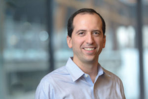
Michael Berger, PhD, holds the Elizabeth and Felix Rohatyn Chair and is Associate Director of the Marie-Josée and Henry R. Kravis Center for Molecular Oncology at Memorial Sloan Kettering Cancer Center, a multidisciplinary initiative to promote precision oncology through genomic analysis to guide the diagnosis and treatment of cancer patients. He is also an Associate Attending Geneticist in the Department of Pathology with expertise in cancer genomics, computational biology, and high-throughput DNA sequencing technology. His laboratory is developing experimental and computational methods to characterize the genetic makeup of individual cancers and identify genomic biomarkers of drug response and resistance. As Scientific Director of Clinical NGS in the Molecular Diagnostics Service, he oversees the development and bioinformatics associated with clinical sequencing assays, and he helped lead the development and implementation of MSK-IMPACT, a comprehensive FDA-authorized tumor sequencing panel that been used to profile more than 60,000 tumors from advanced cancer patients at MSK. The resulting data have enabled the characterization of somatic and germline biomarkers across many cancer types and the identification of mutations associated with clonal hematopoiesis. Dr. Berger also led the development of a clinically validated plasma cell-free DNA assay, MSK-ACCESS, which his laboratory is using to explore tumor evolution, acquired drug resistance, and occult metastatic disease. He received his Bachelor’s Degree in Physics from Princeton University and his Ph.D. in Biophysics from Harvard University.
Hosted by: Utkan Demirci, Ph.D.
Sponsored by: The Canary Center & the Department of Radiology
Stanford University – School of Medicine

CEDSS: The First Cell: A new model for cancer research and treatment
Azra Raza, M.D.
Chan Soon-Shiong Professor of Medicine
Director, Myelodysplastic Syndrome Center
Columbia University Medical Center
Location: Zoom
Meeting URL: https://stanford.zoom.us/s/99340345860
Dial: US: +1 650 724 9799 or +1 833 302 1536 (Toll Free)
Meeting ID: 993 4034 5860
Passcode: 711508
ABSTRACT
Cancer research continues to be predicated on a 1970’s model of research and treatment. Despite half a century of intense research, we are failing spectacularly to improve the outcome for patients with advanced disease. Those who are cured continue to be treated mostly with the older strategies (surgery-chemo-radiation). Our contention is that the real solution to the cancer problem is to diagnose cancer early, at the stage of The First Cell. The rapidly evolving technologies are doing much in this area but need to be expanded. We study a pre-leukemic condition called myelodysplastic syndrome (MDS) with the hope that we can detect the first leukemia cells as the disease transforms to acute myeloid leukemia (AML). Towards this end, we have collected blood and bone marrow samples on MDS and AML patients since 1984. Today, our Tissue Repository has more than 60,000 samples. We propose novel methods to identify surrogate markers that can identify the First Cell through studying the serial samples of patients who evolve from MDS to AML.
ABOUT
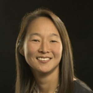
Dr. Raza is a Professor of Medicine and Director of the MDS Center at Columbia University in New York, NY.She started her research in Myelodisplastic Syndromes (MDS) in 1982 and moved to Rush University, Chicago, Illinois in 1992, where she was the Charles Arthur Weaver Professor in Oncology and Director, Division of Myeloid Diseases. The MDS Program, along with a Tissue Repository containing more than 50,000 samples from MDS and acute leukemia patients was successfully relocated to the University of Massachusetts in 2004 and to Columbia University in 2010.
Before moving to New York, Dr. Raza was the Chief of Hematology Oncology and the Gladys Smith Martin Professor of Oncology at the University of Massachussetts in Worcester. She has published the results of her laboratory research and clinical trials in prestigious, peer reviewed journals such as The New England Journal of Medicine, Nature, Blood, Cancer, Cancer Research, British Journal of Hematology, Leukemia, and Leukemia Research. Dr. Raza serves on numerous national and international panels as a reviewer, consultant and advisor and is the recipient of a number of awards.
Hosted by: Utkan Demirci, Ph.D.
Sponsored by: The Canary Center & the Department of Radiology
Stanford University – School of Medicine

Roxana Daneshjou, MD, PhD
Assistant Professor, Biomedical Data Science & Dermatology
Assistant Director, Center of Excellence for Precision Heath & Pharmacogenomics
Director of Informatics, Stanford Skin Innovation and Interventional Research Group
Stanford University
Title: Building Fair and Trustworthy AI for Healthcare
Abstract: AI for healthcare has the potential to revolutionize how we practice medicine. However, to do this in a fair and trustworthy manner requires special attention to how AI models work and their potential biases. In this talk, I will cover the considerations for building AI systems that improve healthcare.

Mildred Cho, PhD
Professor of Pediatrics, Center of Biomedical Ethics
Professor of Medicine, Primary Care and Population Health
Stanford University
Title: Facilitating Patient and Clinician Value Considerations into AI for Precision Medicine
Abstract:
For the development of ethical machine learning (ML) for precision medicine, it is essential to understand how values play into the decision-making process of developers. We conducted five group design exercises with four developer participants each (N=20) who were asked to discuss and record their design considerations in a series of three hypothetical scenarios involving the design of a tool to predict progression to diabetes. In each group, the scenario was first presented as a research project, then as development of a clinical tool for a health care system, and finally as development of a clinical tool for their own health care system. Throughout, developers documented their process considerations using a virtual collaborative whiteboard platform. Our results suggest that developers more often considered client or user perspectives after changing the context of the scenario from research to a tool for a large healthcare setting. Furthermore, developers were more likely to express concerns arising from the patient perspective and societal and ethical issues such as protection of privacy after imagining themselves as patients in the health care system. Qualitative and quantitative data analysis also revealed that developers made reflective/reflexive statements more often in the third round of the design activity (44 times) than in the first (2) or second (6) rounds. These statements included statements on how the activity connected to their real-life work, what they could take away from the exercises and integrate into actual practice, and commentary on being patients within a health care system using AI. These findings suggest that ML developers can be encouraged to link the consequences of their actions to design choices by encouraging “empathy work” that directs them to take perspectives of specific stakeholder groups. This research could inform the creation of educational resources and exercises for developers to better align daily practices with stakeholder values and ethical ML design.

Ifeoma Okoye MBBS, FWACS, FMCR
Professor of Radiology and Director
University of Nigeria Centre for Clinical Trials
College of Medicine, University of Nigeria
Title: Deepening Collaboration with Stanford & Pennsylvania, Toward Developing Joint Strategies to Close the ‘Cancer Care’ & ‘Clinical Trial Volume’ Gap in LMICs
Abstract
In this seminar I will be addressing the dire cancer survival outcomes in low- and middle-income countries (LMICs), with a particular focus on Sub-Saharan Africa. Cancer survival rates in Sub-Saharan Africa are alarmingly low. According to the World Health Organization, cancer deaths in LMICs account for approximately 70% of global cancer fatalities. In Nigeria, the five-year survival rate for breast cancer, one of the most common cancers, stands at a disheartening 10-30%, compared to over 80% in high-income countries. This stark disparity highlights the urgent need for sustained comprehensive cancer interventions in our region.
Here, I will discuss the pivotal role in the cancer control sphere, of a new software, ONCOSEEK, capable of early detecting 11 types of Cancers! It’s particular emphasis on the Patient Perspective, which aligns with our ethos of need for holistic patient care. In addition I will discuss recent developments on collaborative effort with the Gevaert lab at Stanford University and the University of Pennsylvania.

Sophie Ostmeier, MD
Postdoctoral Scholar
Department of Radiology
Stanford School of Medicine
Title: GREEN: Generative Radiology Report Evaluation and Error Notation
Abstract
Evaluating radiology reports is a challenging problem as factual correctness is extremely important due to the need for accurate medical communication about medical images. Existing automatic evaluation metrics either suffer from failing to consider factual correctness (e.g., BLEU and ROUGE) or are limited in their interpretability (e.g., F1CheXpert and F1RadGraph). In this paper, we introduce GREEN (Generative Radiology Report Evaluation and Error Notation), a radiology report generation metric that leverages the natural language understanding of language models to identify and explain clinically significant errors in candidate reports, both quantitatively and qualitatively. Compared to current metrics, GREEN offers: 1) a score aligned with expert preferences, 2) human interpretable explanations of clinically significant errors, enabling feedback loops with end-users, and 3) a lightweight open-source method that reaches the performance of commercial counterparts. We validate our GREEN metric by comparing it to GPT-4, as well as to error counts of 6 experts and preferences of 2 experts. Our method demonstrates not only higher correlation with expert error counts, but simultaneously higher alignment with expert preferences when compared to previous approaches.

Jeong Hoon Lee, PhD
Postdoctoral Researcher
Department of Radiology
Stanford School of Medicine
Title: Leveraging Patch-Level Representation Learning with Vision Transformer for Prostate Cancer Foundation Models
Abstract:
Recent advancements in self-supervised learning (SSL), emerging as an effective approach for imaging foundation models, enable the effective pretraining of AI models across multiple domains without the need for labels. Despite the rapid advancements, their application in medical imaging remains challenging due to the subtle difference between cancer and normal tissue. To address this limitation, in this study, we propose an AI architecture ProViCNet that employs the vision transformer (ViT) based segmentation architecture with patch-level contrastive learning for better feature representation. We validated our model in prostate cancer detection tasks using three types of magnetic resonance imaging (MRI) across multiple centers. To evaluate the performance of feature representation in this model, we performed downstream tasks with respect to Gleason grade score and race prediction. Our model demonstrated significant performance improvements compared to the state-of-the-art segmentation architectures. This study proposes a novel approach to developing foundation models for prostate cancer imaging overcoming SSL limitations.

Ipek Oguz, PhD
Assistant Professor of Computer Science
Assistant Professor of Electrical and Computer Engineering
Assistant Professor of Biomedical Engineering
Vanderbilt University
Title: Medical Image Segmentation and Synthesis
Abstract
Segmentation and synthesis are two fundamental tasks in medical image computing. Segmentation refers to the delineation of the boundaries of a structure of interest in the image, such as an organ, a tumor, or a lesion. Synthesis refers to images created computationally from other data; common examples include cross-modality synthesis and image denoising. This talk will provide an overview of my lab’s recent work in these two broad algorithmic directions in the context of a wide range of medical imaging applications. These driving clinical problems include MR imaging of the brain, OCT imaging of the retina, ultrasound imaging of the placenta, and endoscopic imaging of the kidney. I will also illustrate many problem formulations where synthesis can be used to help segmentation, and vice versa.