“Algorithm Development Lifecycle in Medical Imaging:
Current State and Considerations for the Future”
Luciano M. Prevedello, MD, MPH
Vice-Chair for Medical Informatics and Augmented Intelligence in Imaging
Division Chief, Medical Imaging Informatics
Director, 3D and Advanced Visualization Lab
Associate Professor, Division of Neuroradiology,
Department of Radiology
Ohio State University Wexner Medical Center
Join via Zoom: https://stanford.zoom.us/j/267814863
Abstract:
This presentation will describe some of the most important considerations involved in creating algorithms in medical imaging from inception to deployment as well as continued model improvement and/or monitoring. Examples of experience to date from the OSU laboratory for augmented intelligence in imaging will be provided. New paradigms in model creation and the role of image challenge competitions will also be covered. Current issues with model validation and generalizability will also be introduced as well as considerations for future work in this area.
Refreshments will be provided.

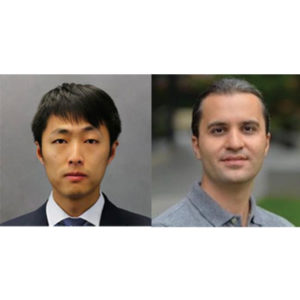
“A Deep Learning Framework for Efficient Registration of MRI and Histopathology Images of the Prostate”
Wei Shao, PhD
Postdoctoral Research Fellow
Department of Radiology
Stanford University
“Applications of Generative Adversarial Networks (GANs) in Medical Imaging”
Saeed Seyyedi, PhD
Paustenbach Research Fellow
Department of Radiology
Stanford University
Join via Zoom: https://stanford.zoom.us/j/593016899
Refreshments will be provided
ABSTRACT (Shao)
Magnetic resonance imaging (MRI) is an increasingly important tool for the diagnosis and treatment of prostate cancer. However, MRI interpretation suffers from high interobserver variability and often misses clinically significant cancers. Registration of histopathology images from patients who have undergone surgical resection of the prostate onto pre-operative MRI images allows direct mapping of cancer location onto MR images. This is essential for the discovery and validation of novel prostate cancer signatures on MRI. Traditional registration approaches can be computationally expensive and require a careful choice of registration hyperparameters. We present a deep learning-based pipeline to accelerate and simplify MRI-histopathology image registration in prostate cancer. Our pipeline consists of preprocessing, transform estimation by deep neural networks, and postprocessing. We refined the registration neural networks, originally trained with 19,642 natural images, by adding 17,821 medical images of the prostate to the training set. The pipeline was evaluated using 99 prostate cancer patients. The addition of the images to the training set significantly (p < 0.001) improved the Dice coefficient and reduced the Hausdorff distance. Our pipeline also achieved comparable accuracy to an existing state-of-the-art algorithm while reducing the computation time from 4.4 minutes to less than 2 seconds.
ABSTRACT (Seyyedi)
Generative adversarial networks (GANs) are advanced types of neural networks where two networks are trained simultaneously to perform two tasks of generation and discrimination. GANs have gained a lot of attention to tackle well known and challenging problems in computer vision applications including medical image analysis tasks such as medical image de-noising, detection and classification, segmentation and reconstruction.In this talk, we will introduce some of the recent advancements of GANs in medical imaging applications and will discuss the recent developments of GAN models to resolve real world imaging challenges.

Ron Kikinis, MD
Director of the Surgical Planning Laboratory
Professor of Radiology
Department of Radiology
Brigham and Women’s Hospital
Harvard Medical School
Title: Evolving Health Care from an Artisanal Organization into an Industrial Enterprise
Refreshments will be provided
Join via Zoom: https://stanford.zoom.us/j/996417088
Abstract: During the last decade, results from basic research in the fields of genetics and immunology have begun to impact treatment in a variety of diseases. Checkpoint therapy, for instance has fundamentally changed the treatment and survival of some patients with melanoma. The medical workplace has transformed from an artisanal organization into an industrial enterprise environment. Workflows in the clinic are increasingly standardized. Their timing and execution are monitored through omnipresent software systems. This has resulted in an acceleration of the pace of care delivery. Imaging and image post-processing have rapidly evolved as well, enabled by ever-increasing computational power, novel sensor systems and novel mathematical approaches. Organizing the data and making it findable and accessible is an ongoing challenge and is investigated through a variety of research efforts. These topics will be reviewed and discussed during the lecture.
About:
Dr. Kikinis is the founding Director of the Surgical Planning Laboratory, Department of Radiology, Brigham and Women’s Hospital, Harvard Medical School, Boston, MA, and a Professor of Radiology at Harvard Medical School. This laboratory was founded in 1990. Before joining Brigham & Women’s Hospital in 1988, he trained as a resident in radiology at the University Hospital in Zurich, and as a researcher in computer vision at the ETH in Zurich, Switzerland. He received his M.D. degree from the University of Zurich, Switzerland, in 1982. In 2004 he was appointed Professor of Radiology at Harvard Medical School. In 2009 he was the inaugural recipient of the MICCAI Society “Enduring Impact Award”. On February 24, 2010 he was appointed the Robert Greenes Distinguished Director of Biomedical Informatics in the Department of Radiology at Brigham and Women’s Hospital. On January 1, 2014, he was appointed “Institutsleiter” of Fraunhofer MEVIS and Professor of Medical Image Computing at the University of Bremen. Since then he is commuting every two months between Bremen and Boston.
During the mid-80’s, Dr. Kikinis developed a scientific interest in image processing algorithms and their use for extracting relevant information from medical imaging data. Due to the explosive increase of both the quantity and complexity of imaging data this area of research is of ever-increasing importance. Dr. Kikinis has led and has participated in research in different areas of science. His activities include technological research (segmentation, registration, visualization, high performance computing), software system development, and biomedical research in a variety of biomedical specialties. The majority of his research is interdisciplinary in nature and is conducted by multidisciplinary teams. The results of his research have been reported in a variety of peer-reviewed journal articles. He is an author and co-author of over 350 peer-reviewed articles.
Follow us on Twitter: @StanfordIBIIS

Tessa Cook, MD, PhD
Assistant Professor of Radiology
Perelman School of Medicine
University of Pennsylvania
Title: Deploying AI in the Clinical Radiology Workflow: Challenges, Opportunities, and Examples
Abstract: Although many radiology AI efforts are focused on pixel-based tasks, there is great potential for AI to impact radiology care delivery and workflow when applied to reports, EMR data, and workflow data. Radiology-pathology correlation, identification of follow-up recommendations, and report segmentation can be used to increase meaningful feedback to radiologists as well as to automate tasks that are currently manual and time-consuming. When deploying AI within the clinical workflow, there are many challenges that may slow down or otherwise affect the integration. Careful consideration of the way in which radiologists may expect to interact with AI results should be undertaken to meaningfully deploy radiology AI in a safe and effective way.

Radiomics and Radio-Genomics: Opportunities for Precision Medicine
Zoom: https://stanford.zoom.us/j/99904033216?pwd=U2tTdUp0YWtneTNUb1E4V2x0OTFMQT09
Pallavi Tiwari, PhD
Assistant Professor of Biomedical Engineering
Associate Member, Case Comprehensive Cancer Center
Director of Brain Image Computing Laboratory
School of Medicine | Case Western Reserve University
Abstract:
In this talk, Dr. Tiwari will focus on her lab’s recent efforts in developing radiomic (extracting computerized sub-visual features from radiologic imaging), radiogenomic (identifying radiologic features associated with molecular phenotypes), and radiopathomic (radiologic features associated with pathologic phenotypes) techniques to capture insights into the underlying tumor biology as observed on non-invasive routine imaging. She will focus on clinical applications of this work for predicting disease outcome, recurrence, progression and response to therapy specifically in the context of brain tumors. She will also discuss current efforts in developing new radiomic features for post-treatment evaluation and predicting response to chemo-radiation treatment. Dr. Tiwari will conclude with a discussion on her lab’s findings in AI + experts, in the context of a clinically challenging problem of post-treatment response assessment on routine MRI scans.
Stanford Molecular Imaging Scholars (SMIS) Program
Quarterly Seminar
Andrew Groll, PhD
Mentor: Craig Levin, PhD
“Initial Experimental Images from a CZT Preclinical PET System”
Brian Lee, PhD
Mentors: Sam Gambhir, MD, PhD; Craig Levin, PhD
“Precision Health Toilet for Cancer Screening”

Stanford Molecular Imaging Scholars (SMIS) Program Quarterly Seminar
Zoom meeting: https://stanford.zoom.us/j/99117388314?pwd=R29OSjlTdUt0a3pLaG5Zc1BFNTJIUT09
Password: 922183
Guolan Lu, PhD
Mentor: Eben Rosenthal, MD; Garry Nolan, PhD
“Co-administered Antibody Improves the Penetration of Antibody-Dye Conjugates into Human Cancers: Implications for AntibodyDrug Conjugates”
Dianna Jeong, PhD
Mentors: Craig Levin, PhD; Shan Wang, PhD
“Novel Detection Approaches for Achieving Ultra-fast time resolution for PET”

Ge Wang, PhD
Clark & Crossan Endowed Chair Professor
Director of the Biomedical Imaging Center
Rensselaer Polytechnic Institute
Troy, New York
Abstract:
AI-based tomography is an important application and a new frontier of machine learning. AI, especially deep learning, has been widely used in computer vision and image analysis, which deal with existing images, improve them, and produce features. Since 2016, deep learning techniques are actively researched for tomography in the context of medicine. Tomographic reconstruction produces images of multi-dimensional structures from externally measured “encoded” data in the form of various transforms (integrals, harmonics, and so on). In this presentation, we provide a general background, highlight representative results, and discuss key issues that need to be addressed in this emerging field.
About:
AI-based X-ray Imaging System (AXIS) lab is led by Dr. Ge Wang, affiliated with the Department of Biomedical Engineering at Rensselaer Polytechnic Institute and the Center for Biotechnology and Interdisciplinary Studies in the Biomedical Imaging Center. AXIS lab focuses on innovation and translation of x-ray computed tomography, optical molecular tomography, multi-scale and multi-modality imaging, and AI/machine learning for image reconstruction and analysis, and has been continuously well funded by federal agencies and leading companies. AXIS group collaborates with Stanford, Harvard, Cornell, MSK, UTSW, Yale, GE, Hologic, and others, to develop theories, methods, software, systems, applications, and workflows.

Join us for a panel on Behavioral XR on Thursday, June 3rd from 9:00 – 10:30 am PDT. The event will start with a one-hour panel discussion featuring Dr. Elizabeth McMahon, a psychologist with a private practice in California; Sarah Hill of Healium, a company developing XR apps for mental fitness based in Missouri; Christian Angern of Sympatient, a company developing VR for anxiety therapy based in Germany; and Marguerite Manteau-Rao of Penumbra, a medical device company based in California. This panel will be moderated by Dr. Walter Greenleaf of Stanford’s Virtual Human Interaction Lab (VHIL) and Dr. Christoph Leuze of the Stanford Medical Mixed Reality (SMMR) program. Immediately following the panel discussion, you are also invited to a 30-minute interactive session with the panelists where questions and ideas can be explored in real time.
Register here to save your place now! After registering, you will receive a confirmation email containing information about joining the meeting.
Please visit this page to subscribe to our events mailing list.
Sponsored by Stanford Medical Mixed Reality (SMMR)

Roxana Daneshjou, MD, PhD
Assistant Professor, Biomedical Data Science & Dermatology
Assistant Director, Center of Excellence for Precision Heath & Pharmacogenomics
Director of Informatics, Stanford Skin Innovation and Interventional Research Group
Stanford University
Title: Building Fair and Trustworthy AI for Healthcare
Abstract: AI for healthcare has the potential to revolutionize how we practice medicine. However, to do this in a fair and trustworthy manner requires special attention to how AI models work and their potential biases. In this talk, I will cover the considerations for building AI systems that improve healthcare.