PHIND Seminar Series: Maintaining Competitive Advantage through Intellectual Capital
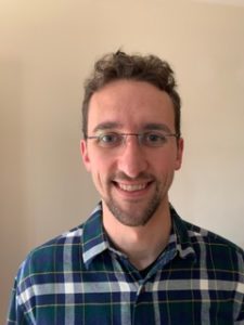
Soody Tronson, M.S., J.D.
Founder
Presque
Location & Timing
11:00am – 12:00pm Seminar & Discussion
Webinar URL: https://stanford.zoom.us/s/98817009379?pwd=U21pYnpCTUlxY3U2TEh4RmhvMXpvQT09
Dial: +1 650 724 9799 or +1 833 302 1536
Webinar ID: 988 1700 9379
Password: 767148
RSVP: https://www.onlineregistrationcenter.com/STronson
ABSTRACT
Digital health has provided a range of solutions, including health and wellness-related management, machine learning algorithms for pattern recognition, AI for genetic analysis, AI-enhanced clinical decision making, and virtual doctors that use AI for patient intake triage. Intellectual property can play an important role in providing a competitive advantage in the digital health industry. We will explore opportunities and challenges for protecting digital health technologies during the program, including patent eligibility challenges and the use of trade secrets.
ABOUT SOODY TRONSON
Soody Tronson is Founding Managing Counsel at STLG Law Firm, counseling domestic and international clients in IP and technology transactions in a wide range of technologies. In 2016 she formed, Presque, a company developing a line of medical devices for mothers and infants.
Soody has over 25 years of operational experience in technology, business, management, and law in start-up and fortune 100 companies, including Schering Plough Pharmaceuticals, Hewlett-Packard Co., Avantec Vascular, and the law firms of HellerEhrman and Townsend and Townsend.
Soody serves in board and leadership capacities with several organizations including the Association of Women in Science STEM to Market national accelerator; Licensing Executives Society USA/Canada, California Lawyers Association, and the Palo Alto Area Bar Association. Soody is a Commissioner with the city of Menlo Park; and an active hands-on volunteer with several civic organizations including Defy Ventures, an entrepreneurship training program for currently and formerly incarcerated. Soody has instructed courses in IP, licensing, and entrepreneurship at the University of California, Stanford University and European institutions. She is the co-author of the book “Women Securing the Future with TIPPSS for IoT: Trust, Identity, Privacy, Protection, Safety, Security for the Internet of Things.”
Hosted by: Garry Gold, MD
Sponsored by the PHIND Center and the Department of Radiology

Judy Gichoya, MD
Assistant Professor
Emory University School of Medicine
Measuring Learning Gains in Man-Machine Assemblage When Augmenting Radiology Work with Artificial Intelligence
Abstract
The work setting of the future presents an opportunity for human-technology partnerships, where a harmonious connection between human-technology produces unprecedented productivity gains. A conundrum at this human-technology frontier remains – will humans be augmented by technology or will technology be augmented by humans? We present our work on overcoming the conundrum of human and machine as separate entities and instead, treats them as an assemblage. As groundwork for the harmonious human-technology connection, this assemblage needs to learn to fit synergistically. This learning is called assemblage learning and it will be important for Artificial Intelligence (AI) applications in health care, where diagnostic and treatment decisions augmented by AI will have a direct and significant impact on patient care and outcomes. We describe how learning can be shared between assemblages, such that collective swarms of connected assemblages can be created. Our work is to demonstrate a symbiotic learning assemblage, such that envisioned productivity gains from AI can be achieved without loss of human jobs.
Specifically, we are evaluating the following research questions: Q1: How to develop assemblages, such that human-technology partnerships produce a “good fit” for visually based cognition-oriented tasks in radiology? Q2: What level of training should pre-exist in the individual human (radiologist) and independent machine learning model for human-technology partnerships to thrive? Q3: Which aspects and to what extent does an assemblage learning approach lead to reduced errors, improved accuracy, faster turn-around times, reduced fatigue, improved self-efficacy, and resilience?
Zoom: https://stanford.zoom.us/j/93580829522?pwd=ZVAxTCtEdkEzMWxjSEQwdlp0eThlUT09

PHIND Seminar Series: Serum Modulation of Mitochondrial Function as a Scalable Sensor of Insulin Resistance
Andrew Lipchik, Ph.D.
Postdoctoral Fellow – Michael Snyder Lab
Stanford University
11:00am – 12:00pm Seminar & Discussion
12:00pm – 12:15pm Reception & Light Refreshments
RSVP: https://stanford.zoom.us/webinar/register/7716009863360/WN_dbeuo7csS8q_AhR88XET0g
Location: Zoom
Webinar URL: . https://stanford.zoom.us/s/96358568342
Webinar ID: 963 5856 8342
Dial: +1 650 724 9799 or +1 833 302 1536 (Toll Free)
Password: 767148
ABSTRACT
The global epidemic of obesity is associated with the dramatic increase in the prevalence of type 2 diabetes mellitus (T2D) with an estimated 400 million people worldwide will have T2D by 2030. T2D is proceeded by insulin resistance (IR) for up to decades prior to onset of T2D. Current estimates suggest approximately one in three individuals are sufficiently insulin resistant to be at risk for IR complications including T2D, coronary heart disease and nonalcoholic fatty liver disease. IR often goes undiagnosed due to the complex, invasive and laborious nature of clamp assays preventing their universal application in the clinic. Surrogate measurements using fasting plasma glucose and insulin levels can estimate IR but are imprecise. There is a need for the identification of new biomarkers and assays for the detection and monitoring of IR. Here, we demonstrate the utility of cellular mitochondrial respiration in response to individuals’ serum as a sensor for personalized monitoring of insulin sensitivity. The modulation of insulin-dependent mitochondrial function by patient serum was highly correlated with insulin sensitivity as determined by the gold-standard modified insulin suppression test (IST). We further applied this methodology to monitor insulin sensitivity over time in response to illness as well as treatment with the insulin sensitizing medication, pioglitazone. Our results demonstrate the development and application of a novel surrogate measurement for the determination and monitoring of insulin sensitivity. This assay offers the advantages of minimal invasiveness and complexity compared to IST as well as superior correlation with IST compared to existing surrogate measurements.
ABOUT ANDREW LIPCHIK
Andrew Lipchik majored in Chemistry at Xavier University where he preformed research on the development of oxygen activation Ni(II) complexes with Dr. Craig Davis and Dr. Michael Baldwin at the University of Cincinnati. He went on to obtain his PhD from Purdue University under mentorship of Dr. Laurie Parker. His thesis work focused on identifying determinants of kinase substrate specificity. This understanding was applied to the development of novel kinase-specific peptide biosensors to monitor intracellular kinase activity. Following his graduate work, he joined the laboratory of Michael Snyder at Stanford University where he has focused on understanding the impact of the immune system on insulin resistance and glucose metabolism.
Hosted by: Garry Gold, M.D.
Sponsored by the PHIND Center and the Department of Radiology

ZOOM LINK HERE
“High Resolution Breast Diffusion Weighted Imaging”
Jessica McKay, PhD
ABSTRACT: Diffusion-weighted imaging (DWI) is a quantitative MRI method that measures the apparent diffusion coefficient (ADC) of water molecules, which reflects cell density and serves as an indication of malignancy. Unfortunately, however, the clinical value of DWI is severely limited by the undesirable features in images that common clinical methods produce, including large geometric distortions, ghosting and chemical shift artifacts, and insufficient spatial resolution. Thus, in order to exploit information encoded in diffusion characteristics and fully assess the clinical value of ADC measurements, it is first imperative to achieve technical advancements of DWI.
In this talk, I will largely focus on the background of breast DWI, providing the clinical motivation for this work and explaining the current standard in breast DWI and alternatives proposed throughout the literature. I will also present my PhD dissertation work in which a novel strategy for high resolution breast DWI was developed. The purpose of this work is to improve DWI methods for breast imaging at 3 Tesla to robustly provide diffusion-weighted images and ADC maps with anatomical quality and resolution. This project has two major parts: Nyquist ghost correction and the use of simultaneous multislice imaging (SMS) to achieve high resolution. Exploratory work was completed to characterize the Nyquist ghost in breast DWI, showing that, although the ghost is mostly linear, the three-line navigator is unreliable, especially in the presence of fat. A novel referenceless ghost correction, Ghost/Object minimization was developed that reduced the ghost in standard SE-EPI and advanced SMS. An advanced SMS method with axial reformatting (AR) is presented for high resolution breast DWI. In a reader study, AR-SMS was preferred by three breast radiologists compared to the standard SE-EPI and readout-segmented-EPI.
“Machine-learning Approach to Differentiation of Benign and Malignant Peripheral Nerve Sheath Tumors: A Multicenter Study”
Michael Zhang, MD
ABSTRACT: Clinicoradiologic differentiation between benign and malignant peripheral nerve sheath tumors (PNSTs) is a diagnostic challenge with important management implications. We sought to develop a radiomics classifier based on 900 features extracted from gadolinium-enhanced, T1-weighted MRI, using the Quantitative Imaging Feature Pipeline and the PyRadiomics package. Additional patient-specific clinical variables were recorded. A radiomic signature was derived from least absolute shrinkage and selection operator, followed by gradient boost machine learning. A training and test set were selected randomly in a 70:30 ratio. We further evaluated the performance of radiomics-based classifier models against human readers of varying medical-training backgrounds. Following image pre-processing, 95 malignant and 171 benign PNSTs were available. The final classifier included 21 features and achieved a sensitivity 0.676, specificity 0.882, and area under the curve (AUC) 0.845. Collectively, human readers achieved sensitivity 0.684, specificity 0.742, and AUC 0.704. We concluded that radiomics using routine gadolinium enhanced, T1-weighted MRI sequences and clinical features can aid in the evaluation of PNSTs, particularly by increasing specificity for diagnosing malignancy. Further improvement may be achieved with incorporation of additional imaging sequences.

PHIND Seminar Series: Identifying Microbiome Markers of Progression of Alzheimer’s Disease
Ami Bhatt, M.D., Ph.D.
Assistant Professor of Medicine (Hematology) and of Genetics
Stanford University
Gavin Sherlock, Ph.D.
Associate Professor of Genetics
Stanford University
11:00am – 12:00pm Seminar & Discussion
RSVP: https://stanford.zoom.us/webinar/register/8016040837299/WN_iBOM7R4XQjOPSb20rkUxbw
Location: Zoom Webinar
Webinar URL: https://stanford.zoom.us/s/99730716280
Webinar ID: 997 3071 6280
Dial: +1 650 724 9799 or +1 833 302 1536 (Toll Free)
Password: 767148
ABOUT AMI BHATT
In perpetual awe of how ‘simple’ microbial organisms can perturb complex, multicellular eukaryotic organisms, Ami Bhatt has chosen to dedicate her research program to inspecting, characterizing and dissecting the microbe-human interface. Nowhere is the interaction between hosts and microbes more potentially impactful than in immunocompromised hosts and global settings where infectious and environmental exposures result in drastic and sometimes fatal health consequences.
Ami’s group identifies problems and questions that arise in the course of routine clinical care. Often in collaboration with investigators at Stanford and beyond, the group applies modern genetic, molecular and computational techniques to seek answers to these questions, better understand host-microbe interactions and decipher how perturbation of these interactions may result in human disease phenotypes.
GAVIN SHERLOCK’S RESEARCH INTERESTS
Adaptive Evolution and the Fitness Landscape: When yeast are evolved under various selective pressures in a chemostat, mutations that arise and provide an adaptive advantage will expand within the population. We have pioneered the use of high throughput sequencing to determine the identity of such mutations, as well as to understand the dynamics of the mutations within the populations, and the interactions between the mutations (such as epistasis). Further, we have developed a DNA barcode based lineage tracking system to determine the distribution of fitness effects (DFE) for newly arising beneficial mutations. We have also characterized what we call the genotype-fitness map for beneficial mutations, and have investigated why beneficial mutations provide a positive fitness effect. We are also interested in how beneficial mutations trade-off for different traits, and how those trade-offs constrain adaptive evolution.
Hosted by: Garry Gold, M.D.
Sponsored by the PHIND Center and the Department of Radiology

Ge Wang, PhD
Clark & Crossan Endowed Chair Professor
Director of the Biomedical Imaging Center
Rensselaer Polytechnic Institute
Troy, New York
Abstract:
AI-based tomography is an important application and a new frontier of machine learning. AI, especially deep learning, has been widely used in computer vision and image analysis, which deal with existing images, improve them, and produce features. Since 2016, deep learning techniques are actively researched for tomography in the context of medicine. Tomographic reconstruction produces images of multi-dimensional structures from externally measured “encoded” data in the form of various transforms (integrals, harmonics, and so on). In this presentation, we provide a general background, highlight representative results, and discuss key issues that need to be addressed in this emerging field.
About:
AI-based X-ray Imaging System (AXIS) lab is led by Dr. Ge Wang, affiliated with the Department of Biomedical Engineering at Rensselaer Polytechnic Institute and the Center for Biotechnology and Interdisciplinary Studies in the Biomedical Imaging Center. AXIS lab focuses on innovation and translation of x-ray computed tomography, optical molecular tomography, multi-scale and multi-modality imaging, and AI/machine learning for image reconstruction and analysis, and has been continuously well funded by federal agencies and leading companies. AXIS group collaborates with Stanford, Harvard, Cornell, MSK, UTSW, Yale, GE, Hologic, and others, to develop theories, methods, software, systems, applications, and workflows.

PHIND Seminar Series: Topics Below
Ahmed Metwally, PhD
“Pre-symptomatic detection of COVID-19 via wearables biosensors”
Postdoctoral Scholar – Michael Snyder, PhD Lab
Department of Genetics
Stanford University
Pierre-Alexandre Fournier, MS
“Continuous remote cardiorespiratory and health monitoring using the Hexoskin biometric shirt”
Co-founder and CEO
Hexoskin
Location: Zoom Webinar
Webinar URL: https://stanford.zoom.us/s/98925964231
Dial: +1 650 724 9799 or +1 833 302 1536 (Toll Free)
Webinar ID: 989 2596 4231
Passcode: 298382
11:00am – 12:00pm Seminar & Discussion
RSVP: https://stanford.zoom.us/webinar/register/WN_bruT-pvvQUePuBLqm2SLkQ
Ahmed Metwally Abstract
Wearable devices digitally measuring vital signs have been used for monitoring health and illness onset and have a high potential for real-time monitoring and disease detection. As such, they are potentially useful during public health crises, such as the current COVID-19 global pandemic. In my talk, I’ll discuss how wearables biosensors can be used as a tool to early detect COVID19 onset using physiological and activity data. By using retrospective smartwatch data, we showed that 63% of the COVID-19 cases could be detected before symptom onset in real-time via the occurrence of extreme elevations in resting heart rate relative to the individual baseline. Our findings suggest that consumer wearables may be used for the large-scale real-time detection of respiratory infections, often pre-symptomatically, and provide an approach for managing epidemics using digital tracking and health monitoring.
About Ahmed Metwally
Ahmed Metwally is a postdoctoral scholar in the Snyder lab at Stanford University. Ahmed received his Ph.D. in Bioinformatics/Bioengineering and MS in Computer Science (focused on Deep Learning), both from the University of Illinois at Chicago (UIC) in 2018. He currently works on developing novel machine learning methods for longitudinal multimodal biomedical data fusion (omics and wearable biosensors data) to early detect cardiometabolic diseases and personalize their treatments. Ahmed has received numerous awards, such as NIH Predoctoral Translational Scientist fellowship, ISMB’20 best talk award, Stanford COVID-19 RISE Grant, second-place award at Stanford Health++ Hackathon, and many travel awards NSF, IEEE, ISCB, and UIUC for various educational and scholarly activities.
Pierre-Alexandre Fournier Abstract
The Hexoskin Connected Health platform will be discussed as an example of a biometric shirt validated for use in telehealth and clinical research. Hexoskin has the only clinically validated biometric garment which provides continuous monitoring of numerous and unique physiological parameters. Hexoskin is an enabling technology for telehealth use cases, such as remote patient monitoring, rehab, and detect the onset of illness. Hexoskin offers a unique set of high-resolution biometric data that can continuously monitor activity, sleep, cardiac and respiratory data. Projects in fields such as cardiology, respiratory, behavioral and physiological psychology, biofeedback research, sleep research, and health will be described. The Hexoskin Connected Health Platform provides researchers with accessible solutions such as the Hexoskin Dashboards, Open API, and Apps to manage, visualize, annotate, analyze, and export raw & processed health data. Data extraction tools allow access to the raw data with time series for machine learning and artificial intelligence projects.
About Pierre-Alexandre Fournier
Pierre-Alexandre Fournier is co-founder and CEO of Hexoskin, a Montreal-based company focused on clinical-grade wearable sensors and AI software for health and clinical research. Hexoskin was founded in 2006 and in 2013 released the first iPhone compatible smart clothing for health monitoring, winning several international awards. In 2018 Hexoskin launched a remote health monitoring system for astronauts on the International Space Station. Hexoskin recently reached the milestone of 100 scientific publications. Pierre-Alexandre earned his MASc and his BEng in Electrical Engineering from the Ecole Polytechnique de Montreal and became a lecturer there teaching machine learning. He completed the Harvard Business School HBX Core program with high honors. Pierre-Alexandre is also an advocate for transparency in healthcare, patient empowerment, and healthcare innovation through design.
Hosted by: Angela McIntyre, Executive Director, eWEAR Initiative
Sponsored by: PHIND Center, Department of Radiology, eWEAR Initiative

PHIND Seminar Series: Maternal Trauma History, Attachment Style, and Depression Are Associated with Broad DNA Methylation Signatures in Infants
Thalia Robakis, M.D., Ph.D.
Associate Professor
Department of Psychiatry
Mount Sinai School of Medicine
Location: Zoom
Webinar URL: https://stanford.zoom.us/s/95483174518
Dial: US: +1 650 724 9799 or +1 833 302 1536 (Toll Free)
Webinar ID: 954 8317 4518
Passcode: 179384
11:00am – 12:00pm Seminar & Discussion
RSVP Here
ABSTRACT
Background: The early environment provides many cues to young organisms that guide their development as they mature. Maternal personality and behavior are an important aspect of the environment of the developing human infant. The molecular mechanisms by which these influences are exerted are not well understood. We attempted to identify whether maternal traits could be associated with alterations in DNA methylation patterns in infants.
Methods: 32 women oversampled for history of depression were recruited in pregnancy and provided information on depressive symptoms, attachment style, and history of early life adversity. Buccal cell DNA was obtained from their infants at six months of age for a large-scale analysis of methylation patterns across 5×10⁶ individual CpG dinucleotides, using clustering-based criteria for significance to control for multiple comparisons. Separately, associations between maternal depression, attachment style, and history of adversity and psychobehavioral outcomes in preschool-age children were examined.
Results: Tens of thousands of individual infant CpGs were alternatively methylated in association with each of the three studied maternal traits. Genes implicated in cell-cell communication, developmental patterning, growth, immune function/inflammatory response, and neurotransmission were identified. The result sets were highly coextensive among the three maternal traits, but areas of divergence exhibited intriguing parallels with behavioral outcomes.
Conclusions: Maternal personality traits are an important aspect of the infant environment that shapes offspring development in many ways. Infant genes that are epigenetically modified in reponse to maternal traits are potential candidate mediators for these effects. We have identified a large number of such genes and demonstrated parallels to clinically measurable outcomes in children.
ABOUT
Dr. Robakis is a psychiatrist with clinical and research interests in perinatal mood disorders and in the contribution of early life experiences to adult mental health and illness. She completed her M.D. as well as a Ph.D. in developmental neurobiology at Columbia University’s Medical Scientist Training Program, residency training in psychiatry at Stanford University School of Medicine, and a research fellowship in perinatal mood disorders also at Stanford. She remained on the clinical faculty at Stanford until 2019, when she accepted a position at the Icahn School of Medicine at Mount Sinai, where she is currently Associate Clinical Professor of Psychiatry and Assistant Director of the Women’s Mental Health Program.
Dr. Robakis’ research interests include the effects of early life stress and disordered attachment on risk for psychiatric illness in the perinatal period, on alterations in metabolism and cognition, and on psychobehavioral development in offspring. She is particularly interested in using epigenetic marks to help identify the biological pathways through which early life experiences exert their effects on outcomes in adulthood and intergenerationally.
Hosted by: Garry Gold, M.D.
Sponsored by the PHIND Center and the Department of Radiology

PHIND Seminar Series: Impact of the Veterans Affairs National Abdominal Aortic Screening Program
Manuel Garcia-Toca, M.D.
Clinical Professor of Surgery
Chief, Division of Vascular Surgery
Santa Clara Valley Medical Center (SCVMC)
Oliver O. Aalami, M.D.
Clinical Associate Professor of Surgery, Vascular Surgery
Lucile Packard Children’s Hospital
Location: Zoom
Webinar URL: https://stanford.zoom.us/s/98417624095
Dial: US: +1 650 724 9799 or +1 833 302 1536 (Toll Free)
Webinar ID: 984 1762 4095
Passcode: 111283
11:00am – 12:00pm Seminar & Discussion
RSVP Here
ABSTRACT
Background: The U.S. Federal Government enacted the Screen for Abdominal Aortic Aneurysms Very Efficiently Act in January 2007. Simultaneously, the Department of Veterans Affairs (VA) implemented a more inclusive AAA screening policy for veteran beneficiaries shortly afterwards.
Our study aimed to evaluate the impact of the VA program on AAA detection rate and all-cause mortality compared to a cohort of patients whose aneurysms were identified by other abdominal imaging.
Methods: We identified veterans with an AAA screening study using the two existing Current Procedural Terminology (CPT) codes (G0389 and 76706). In the comparison group, eligible abdominal imaging studies included ultrasound, computed tomography (CT), and magnetic resonance imaging (MRI) queried according to CPT codes between 2001 and 2018.
We used a difference-in-differences regression model to evaluate the change in aneurysm detection rate and all-cause mortality five years before and eleven years after the VA implemented the screening policy in 2007.
We calculated survival estimates after AAA screening or non-screening imaging of patients with or without AAA diagnosis and used multivariate Cox regression model to evaluate mortality in patients with a positive AAA diagnosis adjusting for patient characteristics and comorbidities.
Results: We identified 3.9 million veterans with abdominal imaging, a total of 303,664 of whom were coded has having an AAA US screening between 2007 and 2018. An AAA diagnosis was made in 4.84% of the screening group vs. 1.3% in the non-screening imaging group P<0.001, yet more aneurysms were found with general imaging studies (50,730 vs.15,449) (Fig 1).
On Kaplan-Meier survival analysis, patients with an AAA diagnosis had higher overall mortality than patients who screened normal; patients with aneurysms found with non-screening imaging had the highest mortality, log-rank P<0.001 (Fig 2).
The difference in differences regression analysis, showed that the absolute AAA detection rate was 1.55% higher (95% CI 1.2- 1.8), and the mortality was 13.89 % lower (95% CI 10.18 %-16.66 %) after the introduction of the screening program in 2007.
Multivariate Cox regression analysis in patients with AAA diagnosis (65-74-year-old) demonstrated a significantly lower 5-year mortality [HR 0.45 (95% CI 0.43-0.48)] for patients in the US Screening group P<0.001.
Conclusions: In a nationwide analysis of VA patients, implementation of AAA screening was associated with improved survival and a higher rate of AAA diagnosis. These findings provide further support for this program’s continuation versus defaulting to incidental recognition following other abdominal imaging.
ABOUT MANUEL GARCIA-TOCA
Dr. Garcia-Toca earned his medical degree at the Universidad Anahuac in Mexico 1999. He has a master’s degree in Health Policy from Stanford University.
He received his general surgery training at the Massachusetts General Hospital and Brown University in 2008. He then completed a Vascular Surgery fellowship at Northwestern University in 2010. Dr. Garcia-Toca is board certified in both surgery and vascular surgery.
Dr. Garcia-Toca joined Stanford Vascular Surgery in 2015. He is currently Clinical Professor of Surgery in the Division of Vascular Surgery. Dr. Garcia-Toca had previously served as an Assistant Professor of Surgery at Brown University. Dr. Garcia Toca is a Staff Surgeon at Santa Clara Valley Medical Center in San Jose.
His research interests include new therapeutic strategies and outcomes for the management of vascular trauma, cerebrovascular diseases, dialysis access, aortic dissection and aneurysms.
ABOUT OLIVER O. AALAMI
Dr. Aalami is a Clinical Associate Professor of Vascular & Endovascular Surgery at Stanford University and the Palo Alto VA and serves as the Lead Director of Stanford’s Biodesign for Digital Health. He is the course director for Biodesign for Digital Health, Building for Digital Health and co-founder of the open source project, CardinalKit, developed to support sensor-based mobile research projects. His primary research focuses on clinically validating the sensors in smartphones and smartwatches in patients with cardiovascular disease to further precision health implementation.
Hosted by: Garry Gold, M.D.
Sponsored by the PHIND Center and the Department of Radiology

Targeted violence continues against Black Americans, Asian Americans, and all people of color. The department of radiology diversity committee is running a racial equity challenge to raise awareness of systemic racism, implicit bias and related issues. Participants will be provided a list of resources on these topics such as articles, podcasts, videos, etc., from which they can choose, with the “challenge” of engaging with one to three media sources prior to our session (some videos are as short as a few minutes). Participants will meet in small-group breakout sessions to discuss what they’ve learned and share ideas.
Please reach out to Marta Flory, flory@stanford.edu with questions. For details about the session, including recommended resources and the Zoom link, please reach out to Meke Faaoso at mfaaoso@stanford.edu.