
"The first known victim to check into the hospital in
Kikwit, Zaire, last month was a 36-year-old lab
technician who complained of headache, fever
and diarrhea. Soon, the nuns and hospital staff caring for him noticed
that his
flesh was beginning to bruise and blister, sloughing off like the
skin of an overripe fruit. Days later, blood started oozing from his eyes,
ears, nose and other orifices, and he began vomiting black sludge, the
residue
of internal organs that were literally rotting inside him. Days after
that, he
was dead.
By May 17, the same hideous illness had killed 77
people in Kikwit--nearly two-thirds of them hospital staff. Alerted by
concerned Zairean health officials, the World Health Organization in
Geneva
dispatched a team of tropical-disease experts to Kikwit, a city of 500,000
that
lies 370 miles east of Zaire's capital,
Kinshasa. On May 11, the Centers for Disease Control and Prevention in
Atlanta,
having tested blood samples sent from Zaire, identified the cause of the
outbreak as the
Ebola virus. Scrambling to contain the deadly pathogen,
Zairean authorities set up
roadblocks outside Kikwit, stopping all travel
in and out of the city."
--Peter Piot and Ellen Wallace "AFRICA'S DEADLY VISITOR; TERROR
IMITATES ART AS THE KILLER EBOLA VIRUS MAKES ANOTHER LETHAL APPEARANCE"
from People Magazine
In 1967, a bizarre new filamentous virus was
discovered
when 31 cases of hemorrhagic fever exploded in Marburg, Germany -- it was
named the Marburg virus.
Nine years later, a virus that was
morphologically idential to but antigenically different from Marburg virus
was isolated in Zaire and was named Ebola virus. To endeavor to
understand these deadly viruses, one must first understand the family they
come from. Read on...
 PROFILING FILOVIRIDAE
THE EBOLA
VIRUS
THE
MARBURG VIRUS
PROFILING FILOVIRIDAE
THE EBOLA
VIRUS
THE
MARBURG VIRUS
What's
going on with Filovirus today?
Want to know
more?
PROFILING FILOVIRIDAE

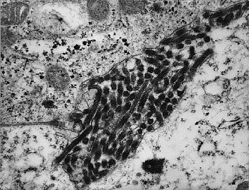
 Filovirus virions are named for their
characteristic threadlike morphology (filo means "filament" in Latin).
With a lipid bilayer envelope encasing a helical nucleocapsid, they are 80
nm in
diameter and have a nucleocapsid length of 800-1000 nm.
Filovirus virions are named for their
characteristic threadlike morphology (filo means "filament" in Latin).
With a lipid bilayer envelope encasing a helical nucleocapsid, they are 80
nm in
diameter and have a nucleocapsid length of 800-1000 nm.
The Filovirus genome is
a single molecule of minus sense ssRNA and is 19 kb in size. This minus
sense genome has seven open reading frames that code for the seven
known structural proteins.
Replication takes place in the
cytoplasm of host cells when the virion removes its
coat and uses its own transcriptase to transcribe its
-ssRNA into the complimentary +ssRNA. Eventually, high
concentrations of replicated viral genomes begin to
appear, marked by the formation of large inclusion
bodies with maturation occurring through budding from the plasma membrane.
 The Filovirus
family was defined only through the morphologic and
replicative mechanisms of the Marburg and Ebola
viruses, compared to other -ssRNA viruses.
Filoviruses are, in fact, known only from a few
isolates in of outbreaks in Africa over the past
years -- including those of the Ebola virus in Zaire,
Sudan, and Ivory Coast, and the Marburg virus in
Zimbabwe and Kenya.
The Filovirus
family was defined only through the morphologic and
replicative mechanisms of the Marburg and Ebola
viruses, compared to other -ssRNA viruses.
Filoviruses are, in fact, known only from a few
isolates in of outbreaks in Africa over the past
years -- including those of the Ebola virus in Zaire,
Sudan, and Ivory Coast, and the Marburg virus in
Zimbabwe and Kenya.
THE
EBOLA VIRUS
The Ebola virus gets its name from a small river in northern Zaire near the
village where the first isolate of the virus was obtained.
**HISTORY**
**TRANSMISSION
and
CLINICAL MANIFESTATIONS**
**VIDEO
CLIPS**
**VACCINE
FOR
EBOLA?**
**MAP
OF ZAIRE**

The Marburg
Virus


The first recorded outbreak of
Marburg virus disease was
in Germany and Yugoslavia in 1967. It was speculated
that the origin of the Marburg virus was Kitmun Cave on
Mount Elgon, along the border between Kenya and Uganda,
because a French expatriate who died in 1980 and a
Danish body who died in 1987 had both visited the cave
before they developed Marburg disease.
When the Marburg virus was first isolated in 1967, these
elongated virus particles already had the reputation of
causing a new disease with high mortality that could
easily be transmitted from patient to caretaker. All
that was known was that the virus was imported from
Uganda with wild-caught African green monkeys and that
it represented a previously unknown group of
viruses.
It was soon discovered that the Marburg and Ebola
viruses belonged to the same family. The Marburg and
Ebola viruses bear many similarities; however, there are
some differences:
They
do not show immunological cross reactivity with each
other.
Ebola
exhibits three transcriptional start and stop codons
while Marburg has just one.
The Ebola
glycoprotein gene produces two transcripts while the
Marburg glycoprotein gene makes one.
What's going on
with Filovirus today?

 Research groups
on the lookout for people or other primates who have
antibodies that can recognise the Marburg virus, Ebola
Zaire virus, and the Reston virus. Click on Badtz-Maru
to know more.
Research groups
on the lookout for people or other primates who have
antibodies that can recognise the Marburg virus, Ebola
Zaire virus, and the Reston virus. Click on Badtz-Maru
to know more.
 1995: Most
recent Ebola outbreak in Kikwit, Zaire. Click on
Pochacco to know more.
1995: Most
recent Ebola outbreak in Kikwit, Zaire. Click on
Pochacco to know more.
 Vaccine
update: Ebola vaccine is years away. Click on Keroppi
to know more.
Vaccine
update: Ebola vaccine is years away. Click on Keroppi
to know more.
NOT satisfied???
Want to know MORE???
Check out
these web sites...

The Big Picture Book of
Viruses: Filoviridae
Ebola -
The Biology of The Virus -
Ebola
Links
EBOLA! Not
just a disease, but a state of mind
Interview
with Dr. Frederick A. Murphy -- by Sean
Henahan


REFERENCES


Oh no! That's the end of my Filovirus
web page...so sad...hope you enjoyed it!
Comments?
Created: February 1, 1999
Last modified:August 4, 1999
In 1967, a bizarre new filamentous virus was
discovered
when 31 cases of hemorrhagic fever exploded in Marburg, Germany -- it was
named the Marburg virus.
Nine years later, a virus that was
morphologically idential to but antigenically different from Marburg virus
was isolated in Zaire and was named Ebola virus. To endeavor to
understand these deadly viruses, one must first understand the family they
come from. Read on...
PROFILING FILOVIRIDAE
THE EBOLA
VIRUS
THE
MARBURG VIRUS
What's
going on with Filovirus today?
Want to know
more?
PROFILING FILOVIRIDAE
Filovirus virions are named for their
characteristic threadlike morphology (filo means "filament" in Latin).
With a lipid bilayer envelope encasing a helical nucleocapsid, they are 80
nm in
diameter and have a nucleocapsid length of 800-1000 nm.
The Filovirus genome is
a single molecule of minus sense ssRNA and is 19 kb in size. This minus
sense genome has seven open reading frames that code for the seven
known structural proteins.
Replication takes place in the
cytoplasm of host cells when the virion removes its
coat and uses its own transcriptase to transcribe its
-ssRNA into the complimentary +ssRNA. Eventually, high
concentrations of replicated viral genomes begin to
appear, marked by the formation of large inclusion
bodies with maturation occurring through budding from the plasma membrane.
The Filovirus
family was defined only through the morphologic and
replicative mechanisms of the Marburg and Ebola
viruses, compared to other -ssRNA viruses.
Filoviruses are, in fact, known only from a few
isolates in of outbreaks in Africa over the past
years -- including those of the Ebola virus in Zaire,
Sudan, and Ivory Coast, and the Marburg virus in
Zimbabwe and Kenya.
The Ebola virus gets its name from a small river in northern Zaire near the village where the first isolate of the virus was obtained.
**HISTORY**
**TRANSMISSION
and
CLINICAL MANIFESTATIONS**
**VIDEO
CLIPS**
**VACCINE
FOR
EBOLA?**
**MAP
OF ZAIRE**
The Marburg
Virus
The first recorded outbreak of
Marburg virus disease was
in Germany and Yugoslavia in 1967. It was speculated
that the origin of the Marburg virus was Kitmun Cave on
Mount Elgon, along the border between Kenya and Uganda,
because a French expatriate who died in 1980 and a
Danish body who died in 1987 had both visited the cave
before they developed Marburg disease.
When the Marburg virus was first isolated in 1967, these
elongated virus particles already had the reputation of
causing a new disease with high mortality that could
easily be transmitted from patient to caretaker. All
that was known was that the virus was imported from
Uganda with wild-caught African green monkeys and that
it represented a previously unknown group of
viruses.
It was soon discovered that the Marburg and Ebola
viruses belonged to the same family. The Marburg and
Ebola viruses bear many similarities; however, there are
some differences:
They
do not show immunological cross reactivity with each
other.
Ebola
exhibits three transcriptional start and stop codons
while Marburg has just one.
The Ebola
glycoprotein gene produces two transcripts while the
Marburg glycoprotein gene makes one.
What's going on
with Filovirus today?
Research groups
on the lookout for people or other primates who have
antibodies that can recognise the Marburg virus, Ebola
Zaire virus, and the Reston virus. Click on Badtz-Maru
to know more.
1995: Most
recent Ebola outbreak in Kikwit, Zaire. Click on
Pochacco to know more.
Vaccine
update: Ebola vaccine is years away. Click on Keroppi
to know more.
NOT satisfied???
Want to know MORE???
Check out
these web sites...
The Big Picture Book of
Viruses: Filoviridae
Ebola -
The Biology of The Virus -
Ebola
Links
EBOLA! Not
just a disease, but a state of mind
Interview
with Dr. Frederick A. Murphy -- by Sean
Henahan
REFERENCES
Oh no! That's the end of my Filovirus
web page...so sad...hope you enjoyed it!
Comments?
Created: February 1, 1999
Last modified:August 4, 1999
They
do not show immunological cross reactivity with each
other. Ebola
exhibits three transcriptional start and stop codons
while Marburg has just one. The Ebola
glycoprotein gene produces two transcripts while the
Marburg glycoprotein gene makes one.
Research groups
on the lookout for people or other primates who have
antibodies that can recognise the Marburg virus, Ebola
Zaire virus, and the Reston virus. Click on Badtz-Maru
to know more. 1995: Most
recent Ebola outbreak in Kikwit, Zaire. Click on
Pochacco to know more.
Vaccine
update: Ebola vaccine is years away. Click on Keroppi
to know more.Check out these web sites...
Ebola - The Biology of The Virus -
Ebola Links
EBOLA! Not just a disease, but a state of mind
Interview with Dr. Frederick A. Murphy -- by Sean Henahan
Comments?
Created: February 1, 1999
Last modified:August 4, 1999