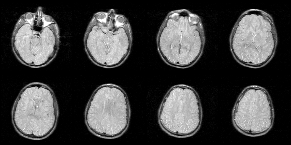
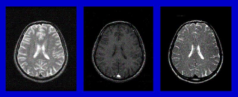
This first set of images is to demonstrate contrast in MRI.
Unlike X-ray, which has only one contrast mechanism, MR imaging
has many different ways of generating contrast. Typically in a
brain scan, at least two different types of MR scan are used, generating
the left two images. All three images are called axial slices, which
means they image a thin horizontal slice of the brain.
Some tissues may show up bright on all images. Others will appear
bright on one image and dark on another. By looking at images with
different types of contrast, doctors can more accurately tell exactly
what kind of tissue they are looking at. In some cases, they can
tell if a tumor is harmful or not.
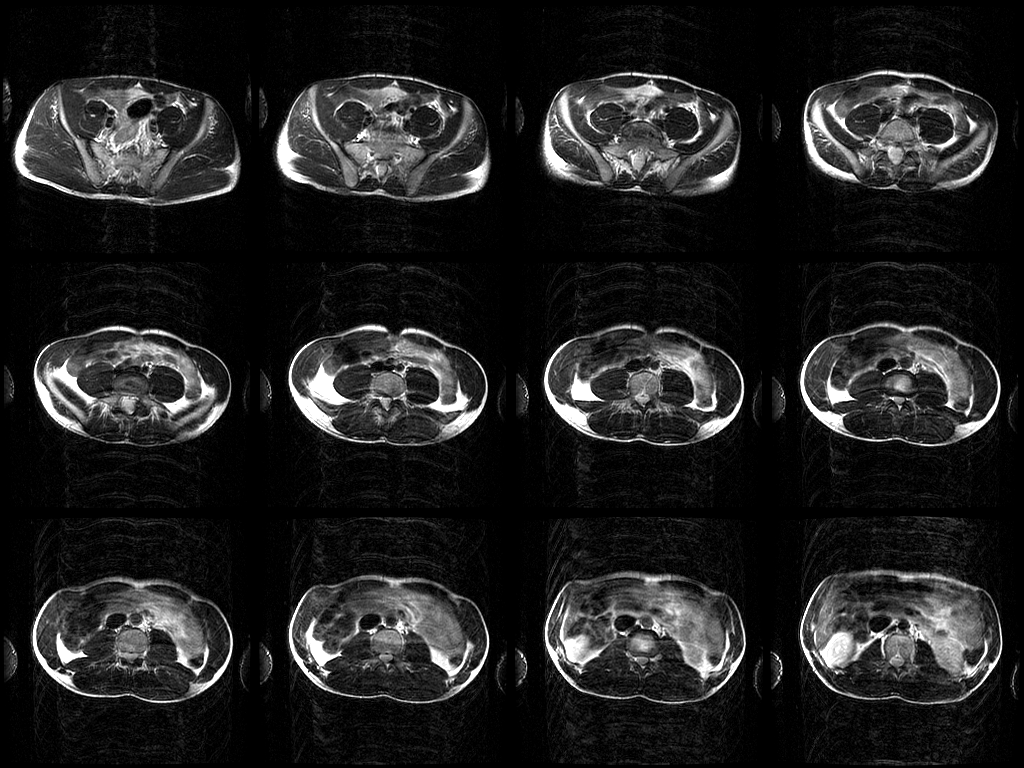
Clinical MRI exams usually include taking images of many slices
through the part of the body being imaged. The set of images below
is a series of saggital (left/right) slices of the brain. Note that
the series starts at one eye and continues toward the other. The "slices"
are about 5mm thick, and their centers are separated by about 10mm.
By collecting a series of slices such as this, doctors have an approximate
representation of a 3-dimensional region of the body -- which increases
the chance of them seeing any problems that exist.
This diagram also shows the directions used to describe images, Inrior-Superior,
Left-Right and Anterior-Posterior.
Click on images below to magnify them...
This is my head! (Inferior -> Superior)
This is my stomach... (Inferior -> Superior) Guess what I ate?!
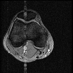
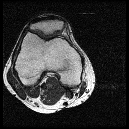
GE Image of knee
My Sequence: Image of knee
Here you can see a knee, with the cap at the top. The
point is to image cartilage between the femur and patella. The
bone is really bright in the right-hand image - I have to work on
this, because in order to have the cartilage brighter, the bone
must be less bright. (For the engineers out there, this is because
it is not possible to form a good image from an FFT if the image-transform
saturates.) In order to reduce the brightness of the bone, a technique
called FAT-SUPRESSION will be used. (Note that the inside of the bone
is actually fatty.)