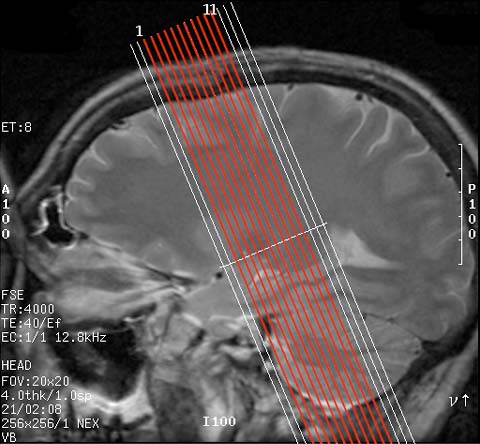
Choose at least 16 slices perpendicular to the long axis of the hippocampus. Always prescribe from anterior to posterior! Here is an example prescription.
Be sure image size is at least 512 x 512, with a FOV of 20 and thickness is as thin as possible, maximum 3.0mm. Goal inplane pixel size is .4 to .5 mm. Goal total volume thickness minimum 4.8 cm.
Doing this will enable you to have a coregistered EPI and structural image. By not separating in time, presumably there is minimal head motion between these two acquisitions. Thus, one can use this single coregistered EPI as a target aligment volume for motion correction. Hence, it is very important that it be acquired very quickly after the hires image. Tell the subject to hold still and scan away.
Make the scan resolution totally coplanar with the structural scan, with the same slice thickenss. Make the inplane resolution as high as possible, at least 128 x 128. Also, make it an even multiple of the structural scan (ie 128 and 512).
Again acquire images at the same resolution as above with your paradigm. Your TR will probably be long, on the order of 3 seconds, and you may not get most of the hippocampal slices. On some scanners, you may have to sacrifice slices to get an acceptable TR.
To select slices, I look at both the highres structurals to see where all the HC is and then at the first EPI grad-echo volume. I usually select slices 3-13. Get as much of the anterior and posterior HC as you can. If you need to sacrifice slices for imaging time, sacrifice those which have little HC, and those obscured by susceptibility artifact.
KEEP TRACK OF WHICH SLICES YOU FUNCTIONALLY IMAGE. You will need this info later.
This can be a useful adjunct to the 2D volume in visualizing the winding collateral sulcus during segmentation, especially for the inexperienced, or for people with obviously complicated collateral sulci.