Dear WMIS trainees, colleagues and friends,
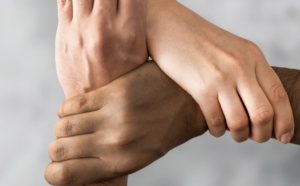
We welcome you to join our upcoming virtual WMIS – Stanford Diversity conference on September 9-11, 2020. We are coming together to reinforce our commitment to diversity and to provide a forum for our team members to engage in meaningful discussions. The conference will provide keynote lectures, scientific presentations and educational lectures from leaders and pioneers in the field, who will discuss important topics related to racial justice, women in STEM and Global Health. We are also offering breakout sessions whereby carefully selected individuals will facilitate a discussion about how to implement more supportive and inclusive practices into our daily professional and personal life. The breakout sessions are designed to enable active involvement of smaller groups where people feel safe to discuss current challenges in the STEM field and actionable solutions.
This conference is free of charge and will provide 9.5 CME credits. Abstracts of all conference presentations and a summary of discussion points and insights provided by all conference participants will be published in Molecular Imaging & Biology. The organizing committee will provide 10 trainee prizes in the form of free WMIS memberships to conference attendants for the 2021 WMIC in Miami.
Website: https://www.wmislive.org

Judy Gichoya, MD
Assistant Professor
Emory University School of Medicine
Measuring Learning Gains in Man-Machine Assemblage When Augmenting Radiology Work with Artificial Intelligence
Abstract
The work setting of the future presents an opportunity for human-technology partnerships, where a harmonious connection between human-technology produces unprecedented productivity gains. A conundrum at this human-technology frontier remains – will humans be augmented by technology or will technology be augmented by humans? We present our work on overcoming the conundrum of human and machine as separate entities and instead, treats them as an assemblage. As groundwork for the harmonious human-technology connection, this assemblage needs to learn to fit synergistically. This learning is called assemblage learning and it will be important for Artificial Intelligence (AI) applications in health care, where diagnostic and treatment decisions augmented by AI will have a direct and significant impact on patient care and outcomes. We describe how learning can be shared between assemblages, such that collective swarms of connected assemblages can be created. Our work is to demonstrate a symbiotic learning assemblage, such that envisioned productivity gains from AI can be achieved without loss of human jobs.
Specifically, we are evaluating the following research questions: Q1: How to develop assemblages, such that human-technology partnerships produce a “good fit” for visually based cognition-oriented tasks in radiology? Q2: What level of training should pre-exist in the individual human (radiologist) and independent machine learning model for human-technology partnerships to thrive? Q3: Which aspects and to what extent does an assemblage learning approach lead to reduced errors, improved accuracy, faster turn-around times, reduced fatigue, improved self-efficacy, and resilience?
Zoom: https://stanford.zoom.us/j/93580829522?pwd=ZVAxTCtEdkEzMWxjSEQwdlp0eThlUT09
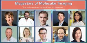
About this Event
This two-day seminar series brings together the brightest minds in molecular imaging to discuss their latest research. Each talk will last 30 min with a full 15 min dedicated for Q&A, related both to their science and their career. So bring that question you’ve always wanted to ask!
Agenda (Note all times are in GMT)
Day 1 (9th November)
13:30 – Ferdia Gallagher (Cambridge University) ‘Clinical imaging of tumour metabolism’
14:15 – David Lewis (CRUK Beatson Institute) ‘Illuminating metabolic vulnerabilities in lung cancer’
15:00 – Federica Pisaneschi (MD Anderson Cancer Center) ‘Imaging ROS burst during myeloid cell activation with 4-[18F]fluoronaphthol’
15:45 – Break
16:00 – Adam Shuhendler (University of Ottawa) ‘Activity-based Sensing by CEST-MRI’
16:45 – Israt Alam (Stanford University) ‘A tale of two biomarkers: visualizing T cell activation with immunoPET’
Day 2 (10th November)
13:30 – André Neves (Cambridge University) ‘Molecular imaging of aberrant glycosylation in cancer’
14:15 – Gilbert Fruhwirth (King’s College London) ‘How non-invasive in vivo cell tracking supports the development of advanced immunotherapeutics’
15:00 – Sarah Bohndiek (Cambridge University) ‘Shedding light on the tumour vasculature’
15:45 – Break
16:00 – John Ronald (Robarts Research Institute) ‘Reporter genes and genome editing for MRI cell tracking: Maybe MRI doesn’t suck?’
16:45 – Michelle James (Stanford University) ‘Shedding light on the immune system to improve detection and treatment of brain diseases using PET’

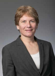
Ge Wang, PhD
Clark & Crossan Endowed Chair Professor
Director of the Biomedical Imaging Center
Rensselaer Polytechnic Institute
Troy, New York
Abstract:
AI-based tomography is an important application and a new frontier of machine learning. AI, especially deep learning, has been widely used in computer vision and image analysis, which deal with existing images, improve them, and produce features. Since 2016, deep learning techniques are actively researched for tomography in the context of medicine. Tomographic reconstruction produces images of multi-dimensional structures from externally measured “encoded” data in the form of various transforms (integrals, harmonics, and so on). In this presentation, we provide a general background, highlight representative results, and discuss key issues that need to be addressed in this emerging field.
About:
AI-based X-ray Imaging System (AXIS) lab is led by Dr. Ge Wang, affiliated with the Department of Biomedical Engineering at Rensselaer Polytechnic Institute and the Center for Biotechnology and Interdisciplinary Studies in the Biomedical Imaging Center. AXIS lab focuses on innovation and translation of x-ray computed tomography, optical molecular tomography, multi-scale and multi-modality imaging, and AI/machine learning for image reconstruction and analysis, and has been continuously well funded by federal agencies and leading companies. AXIS group collaborates with Stanford, Harvard, Cornell, MSK, UTSW, Yale, GE, Hologic, and others, to develop theories, methods, software, systems, applications, and workflows.

Radiology Department-Wide Research Meeting
• Curt Langlotz, MD, PhD: Overview of the AIMI Center
• Brian Hargreaves, PhD: Research Details from Town Hall, Q&A, and COVID19 Updates
Location: Zoom – Details can be found here: https://radresearch.stanford.edu
Meetings will be the 3rd Friday of each month.
Hosted by: Brian Hargreaves, PhD
Sponsored by: the the Department of Radiology
Intersection of Imaging and Therapeutics – MIPS Mini-Retreat
Hosted by: Dr. Heike Daldrup-Link, MD & Dr. Katherine Ferrara, PhD
Sponsored by: Department of Radiology, Molecular Imaging Program at Stanford
The MIPS Mini-retreat series brings together members of the MIPS and greater School of Medicine community to discuss current opportunities for research and collaborations. Each month we will discuss a different topic and we invite all those interested to attend. The mini-retreats will be roughly 1.5-2 hours in length with ample time for discussion throughout. We hope you can join us and spark new collaborations!
Zoom Webinar Information
Webinar URL: https://stanford.zoom.us/j/96227145646
Dial: US: +1 650 724 9799 or +1 833 302 1536 (Toll Free)
Webinar ID: 962 2714 5646
Passcode: 3039816
Agenda (all times are in PST)
8:00-8:05 AM – Opening Remarks – Katherine Ferrara, PhD
8:05-8:20 AM – Image-guided Therapy – Heike Daldrup-Link, MD
8:20-8:35 AM – Imaging Antibody Distribution – Eben Rosenthal, MD
8:35-8:50 AM – MRgFUS and Pancreatic Cancer – Pejman Ghanouni, MD, PhD
8:50-9:05 AM – RefleXion – Lucas Kas Vitzhum, MD
9:05-10:00 AM – Discussion – Moderated by: Katherine Ferrara, PhD
MIPS Seminar Series: Translational Opportunities in Glycoscience
Carolyn Bertozzi, PhD
Director, ChEM-H
Anne T. and Robert M. Bass Professor in the School of Humanities and Sciences
Professor, by courtesy, of Chemical and Systems Biology
Stanford University
Location: Zoom
Webinar URL: . https://stanford.zoom.us/j/94010708043
Dial: US: +1 650 724 9799 or +1 833 302 1536 (Toll Free)
Webinar ID: 940 1070 8043
Passcode: 659236
12:00pm – 12:45pm Seminar & Discussion
RSVP Here
ABSTRACT
Cell surface glycans constitute a rich biomolecular dataset that drives both normal and pathological processes. Their “readers” are glycan-binding receptors that can engage in cell-cell interactions and cell signaling. Our research focuses on mechanistic studies of glycan/receptor biology and applications of this knowledge to new therapeutic strategies. Our recent efforts center on pathogenic glycans in the tumor microenvironment and new therapeutic modalities based on the concept of targeted degradation.
ABOUT
Carolyn Bertozzi is the Baker Family Director of Stanford ChEM-H and the Anne T. and Robert M. Bass Professor of Humanities and Sciences in the Department of Chemistry at Stanford University. She is also an Investigator of the Howard Hughes Medical Institute. Her research focuses on profiling changes in cell surface glycosylation associated with cancer, inflammation and infection, and exploiting this information for development of diagnostic and therapeutic approaches, most recently in the area of immuno-oncology. She is an elected member of the National Academy of Medicine, the National Academy of Sciences, and the American Academy of Arts and Sciences. She also has been awarded the Lemelson-MIT Prize, a MacArthur Foundation Fellowship, the Chemistry for the Future Solvay Prize, among many others.
Hosted by: Katherine Ferrara, PhD
Sponsored by: Molecular Imaging Program at Stanford & the Department of Radiology

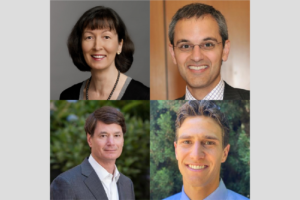
Imaging Immune Cell Modulation – MIPS Mini-Retreat
Hosted by: Dr. Katherine Ferrara, PhD
Sponsored by: Department of Radiology, Molecular Imaging Program at Stanford
The MIPS Mini-retreat series brings together members of the MIPS and greater School of Medicine community to discuss current opportunities for research and collaborations. Each month we will discuss a different topic and we invite all those interested to attend. The mini-retreats will be roughly 1.5-2 hours in length with ample time for discussion throughout. We hope you can join us and spark new collaborations!
Zoom Webinar Information
Webinar URL: https://stanford.zoom.us/j/94969801773
Dial: US: +1 650 724 9799 or +1 833 302 1536 (Toll Free)
Webinar ID: 949 6980 1773
Passcode: 292995
Agenda (all times are in PST)
8:00-8:05 AM – Opening Remarks – Katherine Ferrara, PhD
8:05-8:25 AM – Imaging CAR-T cells – Clinical Need – Crystal Mackall, MD
8:25-8:45 AM – Imaging Immune Cells in Cancer – Clinical Need – Ronald Levy, MD
8:45-9:05 AM – Imaging Approaches – Anna Wu, PhD
9:05-10:00 AM – Discussion – Moderated by: Katherine Ferrara, PhD
Radiology Department-Wide Research Meeting
Location: Zoom – Details can be found here: https://radresearch.stanford.edu
Meetings will be the 3rd Friday of each month.
February 19 Speakers:
Bruce Daniel, MD – Center Overview: IMMERS
Jennifer McNab, PhD – Encoding and Decoding Diffusion MRI
Hosted by: Brian Hargreaves, PhD
Sponsored by: the the Department of Radiology
MIPS Seminar Series: “Convergent, translational research to improve human health”
Joseph M. DeSimone, PhD
Sanjiv Sam Gambhir Professor of Translational Medicine and Chemical Engineering
Departments of Radiology and Chemical Engineering
Graduate School of Business (by Courtesy)
Stanford University
Location: Zoom
Webinar URL: https://stanford.zoom.us/s/98460805010
Dial: +1 650 724 9799 or +1 833 302 1536
Webinar ID: 984 6080 5010
Passcode: 809226
12:00pm – 12:45pm Seminar & Discussion
RSVP Here
ABSTRACT
In many ways, manufacturing processes define what’s possible in society. Central to our interests in the DeSimone laboratory are opportunities to make things using cutting-edge fabrication technologies that can improve human health. This lecture will describe advances in nano- / micro-fabrication and 3D printing technologies that we have made and employed toward this end. Using novel perfluoropolyether materials synthesized in our lab in 2004, we invented the Particle Replication in Non-wetting Templates (PRINT) technology, a high-resolution imprint lithography-based process to fabricate nano- and micro-particles with precise and independent control over particle parameters (e.g. size, shape, modulus, composition, charge, surface chemistry). PRINT brought the precision and uniformity associated with computer industry manufacturing technologies to medicine, resulting in the launch of Liquidia Technologies (NASDAQ: LQDA) and opening new research paths, including to elucidate the influence of specific particle parameters in biological systems (Proc. Natl. Acad. Sci. USA 2008), and to reveal insights to inform the design of vaccines (J. Control. Release 2018), targeted therapeutics (Nano Letters 2015), and even synthetic blood (PNAS 2011). In 2015, we reported the invention of the Continuous Liquid Interface Production (CLIP) 3D printing technology (Science 2015), which overcame major fundamental limitations in polymer 3D printing—slowness, a very limited range of materials, and an inability to create parts with the mechanical and thermal properties needed for widespread, durable utility. By rethinking the physics and chemistry of 3D printing, we created CLIP to eliminate layer-by-layer fabrication altogether. A rapid, continuous process, CLIP generates production-grade parts and is now transforming how products are manufactured in industries including automotive, footwear, and medicine. For example, to help address shortages, CLIP recently enabled a new nasopharyngeal swab for COVID-19 diagnostic testing to go from concept to market in just 20 days, followed by a 400-patient clinical trial at Stanford. Academic laboratories are also using CLIP to pursue new medical device possibilities, including geometrically complex IVRs to optimize drug delivery and implantable chemotherapy absorbers to limit toxic side effects. Vast opportunities exist to use CLIP to pursue next-generation medical devices and prostheses. Moreover, CLIP can improve current approaches; for example, the fabrication of an iontophoretic device we invented several years ago (Sci. Transl. Med. 2015) to drive chemotherapeutics directly into hard-to-reach solid tumors is now being optimized for clinical trials with CLIP. New design opportunities also exist in early detection, for example to improve specimen collection, device performance (e.g. microfluidics, cell sorting, supporting growth and studies with human organoids), and imaging (e.g. PET detectors, ultrasound transducers). Here at Stanford, we are pursuing new 3D printing advances, including software treatment planning for digital therapeutic devices in pediatric medicine, as well as the design of a high-resolution printer capable of single-digit micron resolution to advance microneedle designs as a potent delivery platform for vaccines. The impact of our work on human health ultimately relies on our ability to enable a convergent research program to take shape that allows for new connections to be made among traditionally disparate disciplines and concepts, and to ensure that we maintain a consistent focus on the translational potential of our discoveries and advances.
ABOUT
Joseph M. DeSimone is the Sanjiv Sam Gambhir Professor of Translational Medicine and Chemical Engineering at Stanford University. He holds appointments in the Departments of Radiology and Chemical Engineering with a courtesy appointment in Stanford’s Graduate School of Business. Previously, DeSimone was a professor of chemistry at the University of North Carolina at Chapel Hill and of chemical engineering at North Carolina State University. He is also Co-founder, Board Chair, and former CEO (2014 – 2019) of the additive manufacturing company, Carbon.
DeSimone is responsible for numerous breakthroughs in his career in areas including green chemistry, medical devices, nanomedicine, and 3D printing, also co-founding several companies based on his research. He has published over 350 scientific articles and is a named inventor on over 200 issued patents. Additionally, he has mentored 80 students through Ph.D. completion in his career, half of whom are women and members of underrepresented groups in STEM. In 2016 DeSimone was recognized by President Barack Obama with the National Medal of Technology and Innovation, the highest U.S. honor for achievement and leadership in advancing technological progress. He is also one of only 25 individuals elected to all three branches of the U.S. National Academies (Sciences, Medicine, Engineering). DeSimone received his B.S. in Chemistry in 1986 from Ursinus College and his Ph.D. in Chemistry in 1990 from Virginia Tech.
Hosted by: Katherine Ferrara, PhD
Sponsored by: Molecular Imaging Program at Stanford & the Department of Radiology