
Join us for the 3rd Annual Diversity and Inclusion Forum on Friday, October 9, 2020 on Zoom! This virtual event will highlight innovative workshops developed by our residents and fellows with their educational mentors who have participated in the 2019-2020 cohort of the Leadership Education in Advancing Diversity Program.
The event will be an enriching opportunity for all faculty, residents, fellows, postdocs, students, staff, and community members to learn tools and strategies to enable them to become effective change agents for diversity, equity, and inclusion in medical education.
All are welcome to participate and we look forward to seeing you on Friday, October 9!
Register here:
https://mailchi.mp/046c21726371/diversityforum2020-1632872?e=4a913cab2d
In honor of the 30th anniversary of the Americans with Disabilities Act and October as National Disability Employment Awareness Month, join the Stanford Medicine Abilities Coalition (SMAC) for a first of its kind StanfordMed LIVE event focused on disability. Now more than ever during the COVID-19 pandemic, disabilities, health conditions, and illness impact not only our patients but also all of us, both personally and as members of the Stanford Medicine community. Stanford Medicine leadership will share information, answer questions, and engage in a roundtable discussion about the state of disability at Stanford and how best to support faculty, staff, and students living with disability and chronic illness. We encourage our community to submit questions and comments here to be shared broadly with the Stanford Medicine community. The same link can be used to request any accommodations needed for the livestream. Additional information for the webcast itself will be sent out closer to the event.
Livestream link: https://livestream.com/accounts/1973198/events/9288854

ZOOM LINK HERE
“High Resolution Breast Diffusion Weighted Imaging”
Jessica McKay, PhD
ABSTRACT: Diffusion-weighted imaging (DWI) is a quantitative MRI method that measures the apparent diffusion coefficient (ADC) of water molecules, which reflects cell density and serves as an indication of malignancy. Unfortunately, however, the clinical value of DWI is severely limited by the undesirable features in images that common clinical methods produce, including large geometric distortions, ghosting and chemical shift artifacts, and insufficient spatial resolution. Thus, in order to exploit information encoded in diffusion characteristics and fully assess the clinical value of ADC measurements, it is first imperative to achieve technical advancements of DWI.
In this talk, I will largely focus on the background of breast DWI, providing the clinical motivation for this work and explaining the current standard in breast DWI and alternatives proposed throughout the literature. I will also present my PhD dissertation work in which a novel strategy for high resolution breast DWI was developed. The purpose of this work is to improve DWI methods for breast imaging at 3 Tesla to robustly provide diffusion-weighted images and ADC maps with anatomical quality and resolution. This project has two major parts: Nyquist ghost correction and the use of simultaneous multislice imaging (SMS) to achieve high resolution. Exploratory work was completed to characterize the Nyquist ghost in breast DWI, showing that, although the ghost is mostly linear, the three-line navigator is unreliable, especially in the presence of fat. A novel referenceless ghost correction, Ghost/Object minimization was developed that reduced the ghost in standard SE-EPI and advanced SMS. An advanced SMS method with axial reformatting (AR) is presented for high resolution breast DWI. In a reader study, AR-SMS was preferred by three breast radiologists compared to the standard SE-EPI and readout-segmented-EPI.
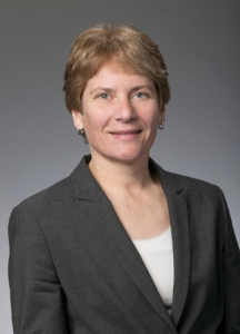
“Machine-learning Approach to Differentiation of Benign and Malignant Peripheral Nerve Sheath Tumors: A Multicenter Study”
Michael Zhang, MD
ABSTRACT: Clinicoradiologic differentiation between benign and malignant peripheral nerve sheath tumors (PNSTs) is a diagnostic challenge with important management implications. We sought to develop a radiomics classifier based on 900 features extracted from gadolinium-enhanced, T1-weighted MRI, using the Quantitative Imaging Feature Pipeline and the PyRadiomics package. Additional patient-specific clinical variables were recorded. A radiomic signature was derived from least absolute shrinkage and selection operator, followed by gradient boost machine learning. A training and test set were selected randomly in a 70:30 ratio. We further evaluated the performance of radiomics-based classifier models against human readers of varying medical-training backgrounds. Following image pre-processing, 95 malignant and 171 benign PNSTs were available. The final classifier included 21 features and achieved a sensitivity 0.676, specificity 0.882, and area under the curve (AUC) 0.845. Collectively, human readers achieved sensitivity 0.684, specificity 0.742, and AUC 0.704. We concluded that radiomics using routine gadolinium enhanced, T1-weighted MRI sequences and clinical features can aid in the evaluation of PNSTs, particularly by increasing specificity for diagnosing malignancy. Further improvement may be achieved with incorporation of additional imaging sequences.

Ge Wang, PhD
Clark & Crossan Endowed Chair Professor
Director of the Biomedical Imaging Center
Rensselaer Polytechnic Institute
Troy, New York
Abstract:
AI-based tomography is an important application and a new frontier of machine learning. AI, especially deep learning, has been widely used in computer vision and image analysis, which deal with existing images, improve them, and produce features. Since 2016, deep learning techniques are actively researched for tomography in the context of medicine. Tomographic reconstruction produces images of multi-dimensional structures from externally measured “encoded” data in the form of various transforms (integrals, harmonics, and so on). In this presentation, we provide a general background, highlight representative results, and discuss key issues that need to be addressed in this emerging field.
About:
AI-based X-ray Imaging System (AXIS) lab is led by Dr. Ge Wang, affiliated with the Department of Biomedical Engineering at Rensselaer Polytechnic Institute and the Center for Biotechnology and Interdisciplinary Studies in the Biomedical Imaging Center. AXIS lab focuses on innovation and translation of x-ray computed tomography, optical molecular tomography, multi-scale and multi-modality imaging, and AI/machine learning for image reconstruction and analysis, and has been continuously well funded by federal agencies and leading companies. AXIS group collaborates with Stanford, Harvard, Cornell, MSK, UTSW, Yale, GE, Hologic, and others, to develop theories, methods, software, systems, applications, and workflows.
MIPS Seminar Series: Translational Opportunities in Glycoscience
Carolyn Bertozzi, PhD
Director, ChEM-H
Anne T. and Robert M. Bass Professor in the School of Humanities and Sciences
Professor, by courtesy, of Chemical and Systems Biology
Stanford University
Location: Zoom
Webinar URL: . https://stanford.zoom.us/j/94010708043
Dial: US: +1 650 724 9799 or +1 833 302 1536 (Toll Free)
Webinar ID: 940 1070 8043
Passcode: 659236
12:00pm – 12:45pm Seminar & Discussion
RSVP Here
ABSTRACT
Cell surface glycans constitute a rich biomolecular dataset that drives both normal and pathological processes. Their “readers” are glycan-binding receptors that can engage in cell-cell interactions and cell signaling. Our research focuses on mechanistic studies of glycan/receptor biology and applications of this knowledge to new therapeutic strategies. Our recent efforts center on pathogenic glycans in the tumor microenvironment and new therapeutic modalities based on the concept of targeted degradation.
ABOUT
Carolyn Bertozzi is the Baker Family Director of Stanford ChEM-H and the Anne T. and Robert M. Bass Professor of Humanities and Sciences in the Department of Chemistry at Stanford University. She is also an Investigator of the Howard Hughes Medical Institute. Her research focuses on profiling changes in cell surface glycosylation associated with cancer, inflammation and infection, and exploiting this information for development of diagnostic and therapeutic approaches, most recently in the area of immuno-oncology. She is an elected member of the National Academy of Medicine, the National Academy of Sciences, and the American Academy of Arts and Sciences. She also has been awarded the Lemelson-MIT Prize, a MacArthur Foundation Fellowship, the Chemistry for the Future Solvay Prize, among many others.
Hosted by: Katherine Ferrara, PhD
Sponsored by: Molecular Imaging Program at Stanford & the Department of Radiology
MIPS Seminar Series: “Convergent, translational research to improve human health”
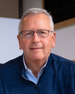
Joseph M. DeSimone, PhD
Sanjiv Sam Gambhir Professor of Translational Medicine and Chemical Engineering
Departments of Radiology and Chemical Engineering
Graduate School of Business (by Courtesy)
Stanford University
Location: Zoom
Webinar URL: https://stanford.zoom.us/s/98460805010
Dial: +1 650 724 9799 or +1 833 302 1536
Webinar ID: 984 6080 5010
Passcode: 809226
12:00pm – 12:45pm Seminar & Discussion
RSVP Here
ABSTRACT
In many ways, manufacturing processes define what’s possible in society. Central to our interests in the DeSimone laboratory are opportunities to make things using cutting-edge fabrication technologies that can improve human health. This lecture will describe advances in nano- / micro-fabrication and 3D printing technologies that we have made and employed toward this end. Using novel perfluoropolyether materials synthesized in our lab in 2004, we invented the Particle Replication in Non-wetting Templates (PRINT) technology, a high-resolution imprint lithography-based process to fabricate nano- and micro-particles with precise and independent control over particle parameters (e.g. size, shape, modulus, composition, charge, surface chemistry). PRINT brought the precision and uniformity associated with computer industry manufacturing technologies to medicine, resulting in the launch of Liquidia Technologies (NASDAQ: LQDA) and opening new research paths, including to elucidate the influence of specific particle parameters in biological systems (Proc. Natl. Acad. Sci. USA 2008), and to reveal insights to inform the design of vaccines (J. Control. Release 2018), targeted therapeutics (Nano Letters 2015), and even synthetic blood (PNAS 2011). In 2015, we reported the invention of the Continuous Liquid Interface Production (CLIP) 3D printing technology (Science 2015), which overcame major fundamental limitations in polymer 3D printing—slowness, a very limited range of materials, and an inability to create parts with the mechanical and thermal properties needed for widespread, durable utility. By rethinking the physics and chemistry of 3D printing, we created CLIP to eliminate layer-by-layer fabrication altogether. A rapid, continuous process, CLIP generates production-grade parts and is now transforming how products are manufactured in industries including automotive, footwear, and medicine. For example, to help address shortages, CLIP recently enabled a new nasopharyngeal swab for COVID-19 diagnostic testing to go from concept to market in just 20 days, followed by a 400-patient clinical trial at Stanford. Academic laboratories are also using CLIP to pursue new medical device possibilities, including geometrically complex IVRs to optimize drug delivery and implantable chemotherapy absorbers to limit toxic side effects. Vast opportunities exist to use CLIP to pursue next-generation medical devices and prostheses. Moreover, CLIP can improve current approaches; for example, the fabrication of an iontophoretic device we invented several years ago (Sci. Transl. Med. 2015) to drive chemotherapeutics directly into hard-to-reach solid tumors is now being optimized for clinical trials with CLIP. New design opportunities also exist in early detection, for example to improve specimen collection, device performance (e.g. microfluidics, cell sorting, supporting growth and studies with human organoids), and imaging (e.g. PET detectors, ultrasound transducers). Here at Stanford, we are pursuing new 3D printing advances, including software treatment planning for digital therapeutic devices in pediatric medicine, as well as the design of a high-resolution printer capable of single-digit micron resolution to advance microneedle designs as a potent delivery platform for vaccines. The impact of our work on human health ultimately relies on our ability to enable a convergent research program to take shape that allows for new connections to be made among traditionally disparate disciplines and concepts, and to ensure that we maintain a consistent focus on the translational potential of our discoveries and advances.
ABOUT
Joseph M. DeSimone is the Sanjiv Sam Gambhir Professor of Translational Medicine and Chemical Engineering at Stanford University. He holds appointments in the Departments of Radiology and Chemical Engineering with a courtesy appointment in Stanford’s Graduate School of Business. Previously, DeSimone was a professor of chemistry at the University of North Carolina at Chapel Hill and of chemical engineering at North Carolina State University. He is also Co-founder, Board Chair, and former CEO (2014 – 2019) of the additive manufacturing company, Carbon.
DeSimone is responsible for numerous breakthroughs in his career in areas including green chemistry, medical devices, nanomedicine, and 3D printing, also co-founding several companies based on his research. He has published over 350 scientific articles and is a named inventor on over 200 issued patents. Additionally, he has mentored 80 students through Ph.D. completion in his career, half of whom are women and members of underrepresented groups in STEM. In 2016 DeSimone was recognized by President Barack Obama with the National Medal of Technology and Innovation, the highest U.S. honor for achievement and leadership in advancing technological progress. He is also one of only 25 individuals elected to all three branches of the U.S. National Academies (Sciences, Medicine, Engineering). DeSimone received his B.S. in Chemistry in 1986 from Ursinus College and his Ph.D. in Chemistry in 1990 from Virginia Tech.
Hosted by: Katherine Ferrara, PhD
Sponsored by: Molecular Imaging Program at Stanford & the Department of Radiology

MIPS Seminar Series: “Circulating Tumor DNA Biomarkers for Therapy Monitoring and Early Detection”
Shan X. Wang, PhD
Leland T. Edwards Professor in the School of Engineering
Professor of Materials Science & Engineering, jointly of Electrical Engineering, and by courtesy of Radiology (Stanford School of Medicine)
Director, Stanford Center for Magnetic Nanotechnology
Stanford University
Location: Zoom
Webinar URL: https://stanford.zoom.us/s/93202777468
Dial: +1 650 724 9799 or +1 833 302 1536
Webinar ID: 932 0277 7468
Passcode: 851144
12:00pm – 12:45pm Seminar & Discussion
RSVP Here
ABSTRACT
Inspired by Dr Sam Gambhir, MIPS, Canary Center, and Stanford CCNE have pursued in vivo imaging and in vitro diagnostic tests for cancer therapeutic response or early detection, respectively, over the last 15+ years. Here I present two successful examples based on circulating tumor DNA (ctDNA) targets in plasma, complementary to imaging modalities such as CT and Ultrasound.
We have developed a simple yet highly sensitive assay for the detection of actionable mutational targets such as Epidermal Growth Factor Receptor (EGFR) and Kirsten rat sarcoma oncogene (KRAS) mutations in the plasma ctDNA from non-small cell lung cancer (NSCLC) patients using giant magnetoresistive (GMR) nanosensors. Our assay achieves lower limits of detection compared to standard fluorescent PCR based assays, and comparable performance to digital PCR methods. In 30 patients with metastatic disease and known EGFR mutation status at diagnosis, our assay achieved 87.5% sensitivity for Exon19 deletion and 90% sensitivity for L858R mutation while retaining 100% specificity; additionally, our assay detected secondary T790M mutation resistance with 96.3% specificity while retaining 100% sensitivity. We re-sampled 13 patients undergoing tyrosine kinase inhibitor (TKI) therapy 2 weeks after initiation to assess response, our GMR assay was 100% accurate in correlation with longitudinal clinical outcome, and the responders identified by the GMR assay had significantly improved progression free survival (PFS) compared to the non-responders. The GMR assay is low cost, rapid, and portable, making it ideal for detecting actionable mutations at diagnosis and non-invasively monitoring treatment response in the clinic.
On another front, we have also developed a highly sensitive and multiplexed assay for the detection of methylated ctDNA targets in plasma samples. Current diagnostic tests for liver cancer in at-risk patients are cumbersome, costly and inaccurate, resulting in a need for accurate blood-based tests. By devising a Layered Analysis of Methylated Biomarkers (LAMB) from the relevant big data, we have discovered a set of DNA targets in the blood that accurately detects liver cancer in these at-risk patients. This set of methylated targets was found by analyzing the genetic information of 3411 liver cancer patients and 1722 healthy people. Our results could lead to clinical adoption of liquid biopsy tests for liver cancer surveillance in high-risk populations and the development of blood tests for other cancers.
ABOUT
Prof. Wang directs the Center for Magnetic Nanotechnology and is a leading expert in biosensors, information storage and spintronics. His research and inventions span across a variety of areas including magnetic biochips, in vitro diagnostics, cancer biomarkers, magnetic nanoparticles, magnetic sensors, magnetoresistive random access memory, and magnetic integrated inductors. He has over 300 publications, and holds 65 issued or pending patents in these and interdisciplinary areas. He was named an inaugural Fred Terman Fellow, and was elected a Fellow of the Institute of Electrical and Electronics Engineers (IEEE) and a Fellow of American Physical Society (APS) for his seminal contributions to magnetic materials and nanosensors. His team won the Grand Challenge Exploration Award from Gates Foundation (2010), the XCHALLENGE Distinguished Award (2014), and the Bold Epic Innovator Award from the XPRIZE Foundation (2017).
Dr. Wang cofounded three high-tech startups in Silicon Valley, including MagArray, Inc. and Flux Biosciences, Inc. In 2018 MagArray launched a first of its kind lung cancer early diagnostic assay based on protein cancer biomarkers and support vector machine (SVM). In 2019, Flux Biosciences launched a human trial to offer at-home testing of fertility based on hormones and magneto-nanosensors. Through his participation in the Center for Cancer Nanotechnology Excellence (as co-PI of the CCNE) and the Joint University Microelectronics Program (JUMP), he is actively engaged in the transformative research of healthcare and is developing emerging memories for energy efficient computing.
Hosted by: Katherine Ferrara, PhD
Sponsored by: Molecular Imaging Program at Stanford & the Department of Radiology

Date: April 10, 2021 (8 AM-6PM)
-
- 8 AM-8:20 AM opening remarks Zainub and Pete
- 8:20 AM-9:20 AM Talk 1 “I fought the law and no one won”
- 10 minute Break
- 9:30 AM-10:30 AM talk 2 students and doctors with disabilities panel
- 20 minute break
-
- 10:50 AM-11:50 AM Breakout
- One hour lunch (TBD)
- 12:50 PM-1:50 PM Talk 3 the frontiers of disability research
- Lisa Meeks is moderating
- Bonnie Swenor invited
- 10 minute break
- 2:00 PM-3:00 PM breakout 2
- 10 minute break
- 3:10 PM-4:10 PM talk 4 do-it-yourself disability advocacy (Poullos/Tolchin with students)
- 4:10 PM-4:30 PM closing remarks
- 4:30 PM-6 PM virtual happy hour

MIPS Seminar Series: Emerging nanophotonic platforms for infectious disease diagnostics: Re-imagining the conventional microbiology toolkit
Jennifer Dionne, PhD
Senior Associate Vice Provost for Research Platforms/Shared Facilities
Associate Professor of Material Science and Engineering and, by courtesy, of Radiology (Molecular Imaging Program at Stanford)
Stanford University
Location: Zoom
Webinar URL: https://stanford.zoom.us/j/95883654314
Dial: +1 650 724 9799 or +1 833 302 1536
Webinar ID: 958 8365 4314
Passcode: 105586
12:00pm – 12:45pm Seminar & Discussion
RSVP Here
ABSTRACT
We present our research controlling light at the nanoscale for infectious disease diagnostics, including detecting bacteria at low concentration, sensing COVID gene sequences, and visualizing in-vivo inter-cellular forces. First, we combine Raman spectroscopy and deep learning to accurately classify bacteria by both species and antibiotic resistance in a single step. We design a convolutional neural network (CNN) for spectral data and train it to identify 30 of the most common bacterial strains from single-cell Raman spectra, achieving antibiotic treatment identification accuracies exceeding 99% and species identification accuracies similar to leading mass spectrometry identification techniques. Our combined Raman-CNN system represents a proof-of-concept for rapid, culture-free identification of bacterial isolates and antibiotic resistance. Second, we describe resonant nanophotonic surfaces, known as “metasurfaces” that enable multiplexed detection of SARS-CoV-2 gene sequences. Our metasurfaces utilize guided mode resonances excited in high refractive index nanostructures. The high quality factor modes produce a large amplification of the electromagnetic field near the nanostructures that increase the response to targeted binding of nucleic acids; simultaneously, the optical signal is beam-steered for multiplexed detection. We describe how this platform can be manufactured at scale for portable, low-cost assays. Finally, we introduce a new class of in vivo optical probes to monitor biological forces with high spatial resolution. Our design is based on upconverting nanoparticles that, when excited in the near-infrared, emit light of a different color and intensity in response to nano-to-microNewton forces. The nanoparticles are sub-30nm in size, do not bleach or photoblink, and can enable deep tissue imaging with minimal tissue autofluorescence. We present the design, synthesis, and characterization of these nanoparticles both in vitro and in vivo, focusing on the forces generated by the roundworm C. elegans as it feeds and digests its bacterial food.
ABOUT
Jennifer Dionne is the Senior Associate Vice Provost of Research Platforms/Shared Facilities and an associate professor of Materials Science and Engineering and, by courtesy, of Radiology at Stanford. She is also an Associate Editor of Nano Letters, director of the DOE-funded Photonics at Thermodynamic Limits Energy Frontier Research Center, and an affiliate faculty of the Wu Tsai Neurosciences Institute, the Institute for Immunity, Transplantation, and Infection, and Bio-X. Jen received her B.S. degrees in Physics and Systems Science and Mathematics from Washington University in St. Louis, her Ph. D. in Applied Physics at the California Institute of Technology in 2009, and her postdoctoral training in Chemistry at Berkeley. Her research develops nanophotonic methods to observe and control chemical and biological processes as they unfold with nanometer scale resolution, emphasizing critical challenges in global health and sustainability. Her work has been recognized with the Alan T. Waterman Award, a NIH Director’s New Innovator Award, a Moore Inventor Fellowship, the Materials Research Society Young Investigator Award, and the Presidential Early Career Award for Scientists and Engineers, and was featured on Oprah’s list of “50 Things that will make you say ‘Wow’!”. Beyond the lab, Jen enjoys exploring the intersection of art and science, long-distance cycling, and reliving her childhood with her two young sons.
Hosted by: Katherine Ferrara, PhD
Sponsored by: Molecular Imaging Program at Stanford & the Department of Radiology

Targeted violence continues against Black Americans, Asian Americans, and all people of color. The department of radiology diversity committee is running a racial equity challenge to raise awareness of systemic racism, implicit bias and related issues. Participants will be provided a list of resources on these topics such as articles, podcasts, videos, etc., from which they can choose, with the “challenge” of engaging with one to three media sources prior to our session (some videos are as short as a few minutes). Participants will meet in small-group breakout sessions to discuss what they’ve learned and share ideas.
Please reach out to Marta Flory, flory@stanford.edu with questions. For details about the session, including recommended resources and the Zoom link, please reach out to Meke Faaoso at mfaaoso@stanford.edu.