Mini-Grand Rounds: Aftershocks: The Coronavirus Pandemic and The New World Disorder
Colin H. Kahl
Senior Fellow at the Freeman Spogli Institute for International Studies
Steven C. Házy Senior Fellow at the Center for International Security and Cooperation
Professor, by courtesy, of Political Science
Co-director of the Center for International Security and Cooperation
7:00am – 7:30am, Zoom
The Stanford Radiology Mini-Grand Round live session events are by invitation only. Invites with link to Zoom video will be sent via email to Department faculty and staff only. Recordings will be made available to the public shortly after the event.

Please note this seminar is now cancelled and will be rescheduled for a future date. Please contact Ashley Williams (ashleylw@stanford.edu) with any questions or concerns. Thank you for your understanding!
IMAGinING THE FUTURE: “Journey Through Academia, Government and Industry: Lessons Learned”
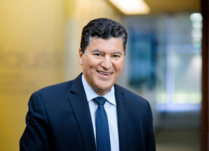
Elias Zerhouni, M.D.
Professor Emeritus
John Hopkins University

Mini-Grand Rounds: The short-run challenges and long-run opportunities of working from home
Nicholas Bloom, PhD
Professor (by courtesy), Economics
Senior Fellow, Stanford Institute for Economic Policy Research
7:00am – 7:30am, Zoom
The Stanford Radiology Mini-Grand Round live session events are by invitation only. Invites with link to Zoom video will be sent via email to Department faculty and staff only. Recordings will be made available to the public shortly after the event.

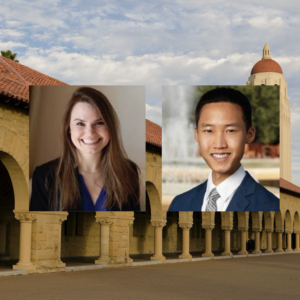
“Tumor-Immune Interactions in TNBC Brain Metastases”
Maxine Umeh Garcia, PhD
ABSTRACT: It is estimated that metastasis is responsible for 90% of cancer deaths, with 1 in every 2 advanced staged triple-negative breast cancer patients developing brain metastases – surviving as little as 4.9 months after metastatic diagnosis. My project hypothesizes that the spatial architecture of the tumor microenvironment reflects distinct tumor-immune interactions that are driven by receptor-ligand pairing; and that these interactions not only impact tumor progression in the brain, but also prime the immune system (early on) to be tolerant of disseminated cancer cells permitting brain metastases. The main goal of my project is to build a model that recapitulates tumor-immune interactions in brain-metastatic triple-negative breast cancer, and use this model to identify novel druggable targets to improve survival outcomes in patients with devastating brain metastases.
“Classification of Malignant and Benign Peripheral Nerve Sheath Tumors With An Open Source Feature Selection Platform”
Michael Zhang, MD
ABSTRACT: Radiographic differentiation of malignant peripheral nerve sheath tumors (MPNSTs) from benign PNSTs is a diagnostic challenge. The former is associated with a five-year survival rate of 30-50%, and definitive management requires gross total surgical with wide negative margins in areas of sensitive neurologic function. This presentation describes a radiomics approach to pre-operatively identifying a diagnosis, thereby possibly avoiding surgical complexity and debilitating symptoms. Using an open-source, feature extraction platform and machine learning, we produce a radiographic signature for MPNSTs based on routine MRI.

Mini-Grand Rounds: Stanford University Medical Center and COVID-19: A Chest Radiologist’s Perspective
Ann Leung, MD
Associate Chair, Clinical Affairs
Professor, Radiology
7:00am – 7:30am, Zoom
The Stanford Radiology Mini-Grand Round live session events are by invitation only. Invites with link to Zoom video will be sent via email to Department faculty and staff only. Recordings will be made available to the public shortly after the event.

Mini-Grand Rounds: The Outlook for Radiology in the Next Phases of the Pandemic and Beyond
David Larson, MD, MBA
Vice Chair, Education and Clinical Operations
Associate Professor, Radiology
7:00am – 7:30am, Zoom
The Stanford Radiology Mini-Grand Round live session events are by invitation only. Invites with link to Zoom video will be sent via email to Department faculty and staff only. Recordings will be made available to the public shortly after the event.

CEDSS: “Multicancer detection of early-stage cancers with simultaneous tissue localization using a plasma cfDNA-based targeted methylation assay”
Eric Fung, M.D., Ph.D.
Senior Medical Director
GRAIL, Inc.
Please see zoom details below:
Meeting URL: https://stanford.zoom.us/j/230531527
Dial: +1 650 724 9799 (US, Canada, Caribbean Toll) or +1 833 302 1536 (US, Canada, Caribbean Toll Free)
Meeting ID: 230 531 527
ABOUT
Dr. Eric Fung is Vice President, Clinical Development at GRAIL, where he leads several clinical development programs in support of the development of a blood-based multi-cancer detection test. Dr. Fung has previously held clinical development and R&D leadership roles at Affymetrix, Vermillion, Ciphergen, and Roche Molecular Diagnostics. Dr. Fung has led clinical trials leading to FDA clearance of multiple IVD products. Dr. Fung received his MD, PhD from the Johns Hopkins University School of Medicine.
Hosted by: Sanjiv Sam Gambhir, M.D., Ph.D.
Sponsored by the Canary Center & the Department of Radiology
Stanford University – School of Medicine

Dear WMIS trainees, colleagues and friends,
We welcome you to join our upcoming virtual WMIS – Stanford Diversity conference on September 9-11, 2020. We are coming together to reinforce our commitment to diversity and to provide a forum for our team members to engage in meaningful discussions. The conference will provide keynote lectures, scientific presentations and educational lectures from leaders and pioneers in the field, who will discuss important topics related to racial justice, women in STEM and Global Health. We are also offering breakout sessions whereby carefully selected individuals will facilitate a discussion about how to implement more supportive and inclusive practices into our daily professional and personal life. The breakout sessions are designed to enable active involvement of smaller groups where people feel safe to discuss current challenges in the STEM field and actionable solutions.
This conference is free of charge and will provide 9.5 CME credits. Abstracts of all conference presentations and a summary of discussion points and insights provided by all conference participants will be published in Molecular Imaging & Biology. The organizing committee will provide 10 trainee prizes in the form of free WMIS memberships to conference attendants for the 2021 WMIC in Miami.
Website: https://www.wmislive.org

CEDSS: “The Origins and Detection of Lethal Prostate Cancer”
Paul Boutros, Ph.D., M.B.A.
Director, Cancer Data Sciences
UCLA
Please see zoom details below:
Meeting URL: https://stanford.zoom.us/s/93515779500
Dial: +1 650 724 9799 or +1 833 302 1536
Meeting ID: 935 1577 9500
Meeting Passcode: 767148
ABOUT
Boutros earned his B.Sc. degree from the University of Waterloo in Chemistry in 2004, and his Ph.D. degree from the University of Toronto, Canada, in Medical Biophysics in 2008. At Toronto, he also earned an executive M.B.A. from the Rothman School of Management. In 2008, Boutros started his independent research career at the Ontario Institute for Cancer Research first as a fellow (2008–2010) and then as principal investigator (2010–2018). He moved to California to join the UCLA faculty in 2018.
Hosted by: Utkan Demirci, Ph.D.
Sponsored by the Canary Center & the Department of Radiology
Stanford University – School of Medicine

ZOOM LINK HERE
“High Resolution Breast Diffusion Weighted Imaging”
Jessica McKay, PhD
ABSTRACT: Diffusion-weighted imaging (DWI) is a quantitative MRI method that measures the apparent diffusion coefficient (ADC) of water molecules, which reflects cell density and serves as an indication of malignancy. Unfortunately, however, the clinical value of DWI is severely limited by the undesirable features in images that common clinical methods produce, including large geometric distortions, ghosting and chemical shift artifacts, and insufficient spatial resolution. Thus, in order to exploit information encoded in diffusion characteristics and fully assess the clinical value of ADC measurements, it is first imperative to achieve technical advancements of DWI.
In this talk, I will largely focus on the background of breast DWI, providing the clinical motivation for this work and explaining the current standard in breast DWI and alternatives proposed throughout the literature. I will also present my PhD dissertation work in which a novel strategy for high resolution breast DWI was developed. The purpose of this work is to improve DWI methods for breast imaging at 3 Tesla to robustly provide diffusion-weighted images and ADC maps with anatomical quality and resolution. This project has two major parts: Nyquist ghost correction and the use of simultaneous multislice imaging (SMS) to achieve high resolution. Exploratory work was completed to characterize the Nyquist ghost in breast DWI, showing that, although the ghost is mostly linear, the three-line navigator is unreliable, especially in the presence of fat. A novel referenceless ghost correction, Ghost/Object minimization was developed that reduced the ghost in standard SE-EPI and advanced SMS. An advanced SMS method with axial reformatting (AR) is presented for high resolution breast DWI. In a reader study, AR-SMS was preferred by three breast radiologists compared to the standard SE-EPI and readout-segmented-EPI.
“Machine-learning Approach to Differentiation of Benign and Malignant Peripheral Nerve Sheath Tumors: A Multicenter Study”
Michael Zhang, MD
ABSTRACT: Clinicoradiologic differentiation between benign and malignant peripheral nerve sheath tumors (PNSTs) is a diagnostic challenge with important management implications. We sought to develop a radiomics classifier based on 900 features extracted from gadolinium-enhanced, T1-weighted MRI, using the Quantitative Imaging Feature Pipeline and the PyRadiomics package. Additional patient-specific clinical variables were recorded. A radiomic signature was derived from least absolute shrinkage and selection operator, followed by gradient boost machine learning. A training and test set were selected randomly in a 70:30 ratio. We further evaluated the performance of radiomics-based classifier models against human readers of varying medical-training backgrounds. Following image pre-processing, 95 malignant and 171 benign PNSTs were available. The final classifier included 21 features and achieved a sensitivity 0.676, specificity 0.882, and area under the curve (AUC) 0.845. Collectively, human readers achieved sensitivity 0.684, specificity 0.742, and AUC 0.704. We concluded that radiomics using routine gadolinium enhanced, T1-weighted MRI sequences and clinical features can aid in the evaluation of PNSTs, particularly by increasing specificity for diagnosing malignancy. Further improvement may be achieved with incorporation of additional imaging sequences.