
Please note this seminar is now cancelled and will be rescheduled for a future date. Please contact Ashley Williams (ashleylw@stanford.edu) with any questions or concerns. Thank you for your understanding!
CEDSS: “The First Cell and the Human Cost of going after Cancer’s last”
Chan Soon-Shiong Professor of Medicine
Director, Myelodysplastic Syndrome Center
Columbia University Medical Center
“Tumor-Immune Interactions in TNBC Brain Metastases”
Maxine Umeh Garcia, PhD
ABSTRACT: It is estimated that metastasis is responsible for 90% of cancer deaths, with 1 in every 2 advanced staged triple-negative breast cancer patients developing brain metastases – surviving as little as 4.9 months after metastatic diagnosis. My project hypothesizes that the spatial architecture of the tumor microenvironment reflects distinct tumor-immune interactions that are driven by receptor-ligand pairing; and that these interactions not only impact tumor progression in the brain, but also prime the immune system (early on) to be tolerant of disseminated cancer cells permitting brain metastases. The main goal of my project is to build a model that recapitulates tumor-immune interactions in brain-metastatic triple-negative breast cancer, and use this model to identify novel druggable targets to improve survival outcomes in patients with devastating brain metastases.
“Classification of Malignant and Benign Peripheral Nerve Sheath Tumors With An Open Source Feature Selection Platform”
Michael Zhang, MD
ABSTRACT: Radiographic differentiation of malignant peripheral nerve sheath tumors (MPNSTs) from benign PNSTs is a diagnostic challenge. The former is associated with a five-year survival rate of 30-50%, and definitive management requires gross total surgical with wide negative margins in areas of sensitive neurologic function. This presentation describes a radiomics approach to pre-operatively identifying a diagnosis, thereby possibly avoiding surgical complexity and debilitating symptoms. Using an open-source, feature extraction platform and machine learning, we produce a radiographic signature for MPNSTs based on routine MRI.
Radiomics and Radio-Genomics: Opportunities for Precision Medicine
Zoom: https://stanford.zoom.us/j/99904033216?pwd=U2tTdUp0YWtneTNUb1E4V2x0OTFMQT09
Pallavi Tiwari, PhD
Assistant Professor of Biomedical Engineering
Associate Member, Case Comprehensive Cancer Center
Director of Brain Image Computing Laboratory
School of Medicine | Case Western Reserve University
Abstract:
In this talk, Dr. Tiwari will focus on her lab’s recent efforts in developing radiomic (extracting computerized sub-visual features from radiologic imaging), radiogenomic (identifying radiologic features associated with molecular phenotypes), and radiopathomic (radiologic features associated with pathologic phenotypes) techniques to capture insights into the underlying tumor biology as observed on non-invasive routine imaging. She will focus on clinical applications of this work for predicting disease outcome, recurrence, progression and response to therapy specifically in the context of brain tumors. She will also discuss current efforts in developing new radiomic features for post-treatment evaluation and predicting response to chemo-radiation treatment. Dr. Tiwari will conclude with a discussion on her lab’s findings in AI + experts, in the context of a clinically challenging problem of post-treatment response assessment on routine MRI scans.
Stanford Molecular Imaging Scholars (SMIS) Program
Quarterly Seminar
Andrew Groll, PhD
Mentor: Craig Levin, PhD
“Initial Experimental Images from a CZT Preclinical PET System”
Brian Lee, PhD
Mentors: Sam Gambhir, MD, PhD; Craig Levin, PhD
“Precision Health Toilet for Cancer Screening”

CEDSS: “Multicancer detection of early-stage cancers with simultaneous tissue localization using a plasma cfDNA-based targeted methylation assay”
Eric Fung, M.D., Ph.D.
Senior Medical Director
GRAIL, Inc.
Please see zoom details below:
Meeting URL: https://stanford.zoom.us/j/230531527
Dial: +1 650 724 9799 (US, Canada, Caribbean Toll) or +1 833 302 1536 (US, Canada, Caribbean Toll Free)
Meeting ID: 230 531 527
ABOUT
Dr. Eric Fung is Vice President, Clinical Development at GRAIL, where he leads several clinical development programs in support of the development of a blood-based multi-cancer detection test. Dr. Fung has previously held clinical development and R&D leadership roles at Affymetrix, Vermillion, Ciphergen, and Roche Molecular Diagnostics. Dr. Fung has led clinical trials leading to FDA clearance of multiple IVD products. Dr. Fung received his MD, PhD from the Johns Hopkins University School of Medicine.
Hosted by: Sanjiv Sam Gambhir, M.D., Ph.D.
Sponsored by the Canary Center & the Department of Radiology
Stanford University – School of Medicine
Stanford Molecular Imaging Scholars (SMIS) Program Quarterly Seminar
Zoom meeting: https://stanford.zoom.us/j/99117388314?pwd=R29OSjlTdUt0a3pLaG5Zc1BFNTJIUT09
Password: 922183
Guolan Lu, PhD
Mentor: Eben Rosenthal, MD; Garry Nolan, PhD
“Co-administered Antibody Improves the Penetration of Antibody-Dye Conjugates into Human Cancers: Implications for AntibodyDrug Conjugates”
Dianna Jeong, PhD
Mentors: Craig Levin, PhD; Shan Wang, PhD
“Novel Detection Approaches for Achieving Ultra-fast time resolution for PET”
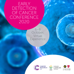
Cancer Research UK, OHSU Knight Cancer Institute and the Canary Center at Stanford, present the Early Detection of Cancer Conference series. The annual Conference brings together experts in early detection from multiple disciplines to share ground breaking research and progress in the field.
The Conference is part of a long-term commitment to invest in early detection research, to understand the biology behind early stage cancers, find new detection and screening methods, and enhance uptake and accuracy of screening.
The 2020 conference will take place October 6-8 virtually.
Cancer Research UK, OHSU Knight Cancer Institute and the Canary Center at Stanford, have been closely monitoring developments relating to the coronavirus (COVID-19) outbreak and reviewing guidance from government bodies. After careful consideration, we have made the decision to convert the Early Detection of Cancer Conference 2020 to a virtual conference, instead of the scheduled in-person conference on October 6-8 in London, UK.
For more information visit the website: http://earlydetectionresearch.com/
CEDSS: “The Origins and Detection of Lethal Prostate Cancer”
Paul Boutros, Ph.D., M.B.A.
Director, Cancer Data Sciences
UCLA
Please see zoom details below:
Meeting URL: https://stanford.zoom.us/s/93515779500
Dial: +1 650 724 9799 or +1 833 302 1536
Meeting ID: 935 1577 9500
Meeting Passcode: 767148
ABOUT
Boutros earned his B.Sc. degree from the University of Waterloo in Chemistry in 2004, and his Ph.D. degree from the University of Toronto, Canada, in Medical Biophysics in 2008. At Toronto, he also earned an executive M.B.A. from the Rothman School of Management. In 2008, Boutros started his independent research career at the Ontario Institute for Cancer Research first as a fellow (2008–2010) and then as principal investigator (2010–2018). He moved to California to join the UCLA faculty in 2018.
Hosted by: Utkan Demirci, Ph.D.
Sponsored by the Canary Center & the Department of Radiology
Stanford University – School of Medicine

ZOOM LINK HERE
“High Resolution Breast Diffusion Weighted Imaging”
Jessica McKay, PhD
ABSTRACT: Diffusion-weighted imaging (DWI) is a quantitative MRI method that measures the apparent diffusion coefficient (ADC) of water molecules, which reflects cell density and serves as an indication of malignancy. Unfortunately, however, the clinical value of DWI is severely limited by the undesirable features in images that common clinical methods produce, including large geometric distortions, ghosting and chemical shift artifacts, and insufficient spatial resolution. Thus, in order to exploit information encoded in diffusion characteristics and fully assess the clinical value of ADC measurements, it is first imperative to achieve technical advancements of DWI.
In this talk, I will largely focus on the background of breast DWI, providing the clinical motivation for this work and explaining the current standard in breast DWI and alternatives proposed throughout the literature. I will also present my PhD dissertation work in which a novel strategy for high resolution breast DWI was developed. The purpose of this work is to improve DWI methods for breast imaging at 3 Tesla to robustly provide diffusion-weighted images and ADC maps with anatomical quality and resolution. This project has two major parts: Nyquist ghost correction and the use of simultaneous multislice imaging (SMS) to achieve high resolution. Exploratory work was completed to characterize the Nyquist ghost in breast DWI, showing that, although the ghost is mostly linear, the three-line navigator is unreliable, especially in the presence of fat. A novel referenceless ghost correction, Ghost/Object minimization was developed that reduced the ghost in standard SE-EPI and advanced SMS. An advanced SMS method with axial reformatting (AR) is presented for high resolution breast DWI. In a reader study, AR-SMS was preferred by three breast radiologists compared to the standard SE-EPI and readout-segmented-EPI.
“Machine-learning Approach to Differentiation of Benign and Malignant Peripheral Nerve Sheath Tumors: A Multicenter Study”
Michael Zhang, MD
ABSTRACT: Clinicoradiologic differentiation between benign and malignant peripheral nerve sheath tumors (PNSTs) is a diagnostic challenge with important management implications. We sought to develop a radiomics classifier based on 900 features extracted from gadolinium-enhanced, T1-weighted MRI, using the Quantitative Imaging Feature Pipeline and the PyRadiomics package. Additional patient-specific clinical variables were recorded. A radiomic signature was derived from least absolute shrinkage and selection operator, followed by gradient boost machine learning. A training and test set were selected randomly in a 70:30 ratio. We further evaluated the performance of radiomics-based classifier models against human readers of varying medical-training backgrounds. Following image pre-processing, 95 malignant and 171 benign PNSTs were available. The final classifier included 21 features and achieved a sensitivity 0.676, specificity 0.882, and area under the curve (AUC) 0.845. Collectively, human readers achieved sensitivity 0.684, specificity 0.742, and AUC 0.704. We concluded that radiomics using routine gadolinium enhanced, T1-weighted MRI sequences and clinical features can aid in the evaluation of PNSTs, particularly by increasing specificity for diagnosing malignancy. Further improvement may be achieved with incorporation of additional imaging sequences.

Ge Wang, PhD
Clark & Crossan Endowed Chair Professor
Director of the Biomedical Imaging Center
Rensselaer Polytechnic Institute
Troy, New York
Abstract:
AI-based tomography is an important application and a new frontier of machine learning. AI, especially deep learning, has been widely used in computer vision and image analysis, which deal with existing images, improve them, and produce features. Since 2016, deep learning techniques are actively researched for tomography in the context of medicine. Tomographic reconstruction produces images of multi-dimensional structures from externally measured “encoded” data in the form of various transforms (integrals, harmonics, and so on). In this presentation, we provide a general background, highlight representative results, and discuss key issues that need to be addressed in this emerging field.
About:
AI-based X-ray Imaging System (AXIS) lab is led by Dr. Ge Wang, affiliated with the Department of Biomedical Engineering at Rensselaer Polytechnic Institute and the Center for Biotechnology and Interdisciplinary Studies in the Biomedical Imaging Center. AXIS lab focuses on innovation and translation of x-ray computed tomography, optical molecular tomography, multi-scale and multi-modality imaging, and AI/machine learning for image reconstruction and analysis, and has been continuously well funded by federal agencies and leading companies. AXIS group collaborates with Stanford, Harvard, Cornell, MSK, UTSW, Yale, GE, Hologic, and others, to develop theories, methods, software, systems, applications, and workflows.