
Stanford School of Medicine’s
1st Annual Conference on Disability in Healthcare and Medicine
Saturday, June 20, 2020
8:00am – 2:30pm Pacific Daylight Time (PDT)
Zoom Webinar
The conference goals are:
- Supporting students and healthcare providers with disabilities
- Training healthcare providers to better care for patients with disabilities
- Research into the intersection of providers and patients with disabilities
Target audience:
- Nursing students and nurses
- PA students and PA’s
- Medical students and medical doctors
- All other interested healthcare providers and allies

Thursday MIPS Roundtable: Faculty Lab Showcase
MIPS Roundtables are every other Thursday from 1:30-2:30pm showcasing various topics and are open to all interested.
1:30-2:00 PM | Dr. Brian Rutt, Ph.D.
Cellular & Molecular MRI Laboratory (CMMRIL)
Professor of Radiology
Stanford University
2:00-2:30 PM | Dr. Kathy Ferrara, Ph.D.
Ferrara Laboratory: Image-guided Drug Delivery
Professor of Radiology
Stanford University
Please note Zoom information does change week to week.
6/25 Webinar URL: https://stanford.zoom.us/j/91635637393?pwd=c09vUXYyeU5VeHJBaUJVRHQrT3FJdz09
Dial: +1 650 724 9799 or +1 833 302 1536
Webinar ID: 916 3563 7393
Webinar Password: 271364
Thursday MIPS Roundtable: Meet our MIPS Instructors
MIPS Roundtables are every other Thursday from 1:30-2:30pm showcasing various topics and are open to all interested. Note we will take a break through late July and August.
1:30-2:00 PM | Dr. Ahmed El Kaffas, Ph.D.
Translational Ultrasound for Tissue Characterization and Stimulation
Instructor, Radiology
Stanford University
2:00-2:30 PM | Dr. Brett Fite, Ph.D.
Combining Focal and Immunotherapies
Instructor, Radiology
Stanford University
Please note Zoom information does change week to week.
7/9 Webinar URL: https://stanford.zoom.us/j/91909413178
Dial: +1 650 724 9799 or +1 833 302 1536
Webinar ID: 919 0941 3178
Password: 572746

Thursday MIPS Roundtable: Meet our MIPS Instructors
MIPS Roundtables are Thursdays from 1:30-2:30pm showcasing various topics and are open to all interested. Note this will be our last summer Roundtable and we will take a break through late July and August.
1:30-2:00 PM | Dr. Josquin Foiret, Ph.D.
High throughput ultrasound imaging for improved diagnosis
Instructor, Radiology
Stanford University
2:00-2:30 PM | Dr. Jinghang Xie, Ph.D.
TESLA probes for imaging T cell-mediated cytotoxic response to immunotherapy
Instructor, Radiology
Stanford University
Please note Zoom information does change week to week.
7/16 Webinar URL: https://stanford.zoom.us/j/94952044130
Dial: +1 650 724 9799 or +1 833 302 1536
Webinar ID: 949 5204 4130
Password: 963699
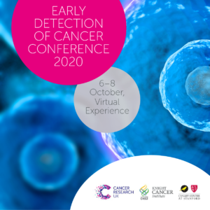
Cancer Research UK, OHSU Knight Cancer Institute and the Canary Center at Stanford, present the Early Detection of Cancer Conference series. The annual Conference brings together experts in early detection from multiple disciplines to share ground breaking research and progress in the field.
The Conference is part of a long-term commitment to invest in early detection research, to understand the biology behind early stage cancers, find new detection and screening methods, and enhance uptake and accuracy of screening.
The 2020 conference will take place October 6-8 virtually.
Cancer Research UK, OHSU Knight Cancer Institute and the Canary Center at Stanford, have been closely monitoring developments relating to the coronavirus (COVID-19) outbreak and reviewing guidance from government bodies. After careful consideration, we have made the decision to convert the Early Detection of Cancer Conference 2020 to a virtual conference, instead of the scheduled in-person conference on October 6-8 in London, UK.
For more information visit the website: http://earlydetectionresearch.com/

Join us for the 3rd Annual Diversity and Inclusion Forum on Friday, October 9, 2020 on Zoom! This virtual event will highlight innovative workshops developed by our residents and fellows with their educational mentors who have participated in the 2019-2020 cohort of the Leadership Education in Advancing Diversity Program.
The event will be an enriching opportunity for all faculty, residents, fellows, postdocs, students, staff, and community members to learn tools and strategies to enable them to become effective change agents for diversity, equity, and inclusion in medical education.
All are welcome to participate and we look forward to seeing you on Friday, October 9!
Register here:
https://mailchi.mp/046c21726371/diversityforum2020-1632872?e=4a913cab2d
CEDSS: “The Origins and Detection of Lethal Prostate Cancer”
Paul Boutros, Ph.D., M.B.A.
Director, Cancer Data Sciences
UCLA
Please see zoom details below:
Meeting URL: https://stanford.zoom.us/s/93515779500
Dial: +1 650 724 9799 or +1 833 302 1536
Meeting ID: 935 1577 9500
Meeting Passcode: 767148
ABOUT
Boutros earned his B.Sc. degree from the University of Waterloo in Chemistry in 2004, and his Ph.D. degree from the University of Toronto, Canada, in Medical Biophysics in 2008. At Toronto, he also earned an executive M.B.A. from the Rothman School of Management. In 2008, Boutros started his independent research career at the Ontario Institute for Cancer Research first as a fellow (2008–2010) and then as principal investigator (2010–2018). He moved to California to join the UCLA faculty in 2018.
Hosted by: Utkan Demirci, Ph.D.
Sponsored by the Canary Center & the Department of Radiology
Stanford University – School of Medicine
In honor of the 30th anniversary of the Americans with Disabilities Act and October as National Disability Employment Awareness Month, join the Stanford Medicine Abilities Coalition (SMAC) for a first of its kind StanfordMed LIVE event focused on disability. Now more than ever during the COVID-19 pandemic, disabilities, health conditions, and illness impact not only our patients but also all of us, both personally and as members of the Stanford Medicine community. Stanford Medicine leadership will share information, answer questions, and engage in a roundtable discussion about the state of disability at Stanford and how best to support faculty, staff, and students living with disability and chronic illness. We encourage our community to submit questions and comments here to be shared broadly with the Stanford Medicine community. The same link can be used to request any accommodations needed for the livestream. Additional information for the webcast itself will be sent out closer to the event.
Livestream link: https://livestream.com/accounts/1973198/events/9288854

ZOOM LINK HERE
“High Resolution Breast Diffusion Weighted Imaging”
Jessica McKay, PhD
ABSTRACT: Diffusion-weighted imaging (DWI) is a quantitative MRI method that measures the apparent diffusion coefficient (ADC) of water molecules, which reflects cell density and serves as an indication of malignancy. Unfortunately, however, the clinical value of DWI is severely limited by the undesirable features in images that common clinical methods produce, including large geometric distortions, ghosting and chemical shift artifacts, and insufficient spatial resolution. Thus, in order to exploit information encoded in diffusion characteristics and fully assess the clinical value of ADC measurements, it is first imperative to achieve technical advancements of DWI.
In this talk, I will largely focus on the background of breast DWI, providing the clinical motivation for this work and explaining the current standard in breast DWI and alternatives proposed throughout the literature. I will also present my PhD dissertation work in which a novel strategy for high resolution breast DWI was developed. The purpose of this work is to improve DWI methods for breast imaging at 3 Tesla to robustly provide diffusion-weighted images and ADC maps with anatomical quality and resolution. This project has two major parts: Nyquist ghost correction and the use of simultaneous multislice imaging (SMS) to achieve high resolution. Exploratory work was completed to characterize the Nyquist ghost in breast DWI, showing that, although the ghost is mostly linear, the three-line navigator is unreliable, especially in the presence of fat. A novel referenceless ghost correction, Ghost/Object minimization was developed that reduced the ghost in standard SE-EPI and advanced SMS. An advanced SMS method with axial reformatting (AR) is presented for high resolution breast DWI. In a reader study, AR-SMS was preferred by three breast radiologists compared to the standard SE-EPI and readout-segmented-EPI.
“Machine-learning Approach to Differentiation of Benign and Malignant Peripheral Nerve Sheath Tumors: A Multicenter Study”
Michael Zhang, MD
ABSTRACT: Clinicoradiologic differentiation between benign and malignant peripheral nerve sheath tumors (PNSTs) is a diagnostic challenge with important management implications. We sought to develop a radiomics classifier based on 900 features extracted from gadolinium-enhanced, T1-weighted MRI, using the Quantitative Imaging Feature Pipeline and the PyRadiomics package. Additional patient-specific clinical variables were recorded. A radiomic signature was derived from least absolute shrinkage and selection operator, followed by gradient boost machine learning. A training and test set were selected randomly in a 70:30 ratio. We further evaluated the performance of radiomics-based classifier models against human readers of varying medical-training backgrounds. Following image pre-processing, 95 malignant and 171 benign PNSTs were available. The final classifier included 21 features and achieved a sensitivity 0.676, specificity 0.882, and area under the curve (AUC) 0.845. Collectively, human readers achieved sensitivity 0.684, specificity 0.742, and AUC 0.704. We concluded that radiomics using routine gadolinium enhanced, T1-weighted MRI sequences and clinical features can aid in the evaluation of PNSTs, particularly by increasing specificity for diagnosing malignancy. Further improvement may be achieved with incorporation of additional imaging sequences.

CEDSS: Systematic identification of fluid-based biomarkers for ovarian and prostate cancer
Thomas Kislinger, Ph.D.
Professor & Chair
Department of Medical Biophysics
University of Toronto
Senior Scientist
Princess Margaret Cancer Centre
Zoom Webinar Details
Meeting URL: https://stanford.zoom.us/s/94878578384
Dial: +1 650 724 9799 or +1 833 302 1536
Webinar ID: 948 7857 8384
Passcode: 692692
Register Here
ABOUT
Thomas Kislinger received his MSc in Analytical Chemistry from the University of Munich, Germany (1998). He completed his PhD in 2001, investigating the role of Advanced Glycation Endproducts in diabetic vascular complications at the University of Erlangen, Germany and Columbia University, New York. Between 2002 and 2006 he completed a post-doctoral fellowship at the University of Toronto. In 2006 he joined the Princess Margaret Cancer Centre as an independent investigator. Dr. Kislinger holds positions as Senior Scientist at the Princess Margaret Cancer Centre and as Professor and Chair at the University of Toronto in the Department of Medical Biophysics. The Kislinger lab applies proteomics technologies to translational and basic cancer biology. This includes the development of novel proteomics methodologies, identification of liquid biopsy signatures and the molecular identification of novel cell surface markers.
Hosted by: Utkan Demirci, Ph.D.
Sponsored by: The Canary Center & the Department of Radiology
Stanford University – School of Medicine