1:00pm-2:00pm: Presentation and Q&A
2:00pm-2:30pm: Light Refreshments
Talk title: Precision Imaging Guides Cancer Therapy
Historically, many of the gains in medicine have been achieved by uniformly applying medical insights to large groups of patients. A combination of increasing biological understanding of the heterogeneity of disease processes and the concurrent expansion of available selective therapeutic interventions has provided the opportunity to improve group outcomes by optimizing treatment on an individual basis. In many cases, molecular imaging is ideally suited to serve as a biomarker to guide therapy selection and dosing, and to provide an early assessment of efficacy. This presentation highlights by example several areas in which imaging helps determine target engagement, dynamic cellular response to treatment, and the effectiveness of immune modulation in oncologic treatment.

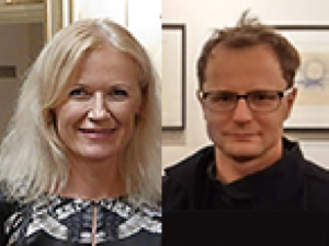
“Messaging in the Age of Microtargeting”
John Stafford
Assistant Vice President
Digital Strategy
Stanford University
Bjorn Carey
Senior Director
Digital Strategy
Stanford University
Join via Zoom: https://stanford.zoom.us/j/400566542
Abstract:
Communications has become increasingly data-driven, targeted, and personalized. This has changed how Stanford analyzes communications opportunities from a research perspective and how it engages with relevant audiences. In this presentation, John and Bjorn will share the data and communications strategy underlying three communications initiatives and the resulting execution. They will also provide practical advice for individual thought leadership and communications in this dynamic environment.
About:
John Stafford, MA ’06, is currently Assistant Vice President for Digital Strategy at Stanford, the most senior digital communications role in the university. John is responsible for all aspects of creating a world-class digital communications function: setting the group’s strategy, building analytics and insight programs, counseling on crisis communications, leading multi-channel messaging initiatives, and advising colleagues across the University. He received a Master’s Degree in Communication from Stanford, a B.A. in History from the University of San Francisco, and was a founding advisor to Stanford Medicine X.
Refreshments will be provided.

MIPS Seminar: “Tiny Bubbles, Big Impact: Exploring applications of nanobubbles in ultrasound molecular imaging and therapy”
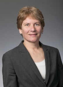
Agata A. Exner, Ph.D.
Professor of Radiology and Biomedical Engineering
Department of Radiology
Case Western Reserve
Location: Beckman Center, B230
2:00pm – 3:00pm Seminar & Discussion
ABSTRACT
Sub-micron shell stabilized gas bubbles (aka nanobubbles (NB) or ultrafine bubbles) have gained momentum as a robust contrast agent for molecular imaging and therapy using ultrasound. The small size, extended stability and high concentration of nanobubbles make them an ideal tool for new applications of contrast enhanced ultrasound and ultra-
sound-mediated therapy, especially in oncology-related problems. Compared to microbub-bles, nanobubbles can provide superior tumor delineation, identify biomarkers on the vascu-lature and on tumors cells and facilitate drug and gene delivery into tumor tissue. The pat-terns of tissue enhancement under nonlinear ultrasound imaging of nanobubbles are distinct from conventional microbubbles especially in tissues exhibiting vascular hyperper-meability. Specifically, NB kinetics, quantified via time intensity curve analysis, typically show a marked delay in the washout rate and significantly increased area under the curve compared to larger bubbles. This effect is further enhanced by molecular targeting to cellular biomarkers, such as the prostate specific membrane antigen (PSMA) or the receptor protein tyrosine phosphatase, PTPmu. The unique contrast enhancement dynamics of nanobubbles are likely to be a result of direct bubble extravasation and prolonged retention of intact bubbles in target tissue. Thus, understanding the underlying mechanisms behind the unique nanobubble behavior can be the driver of significant future innovations in contrast enhanced ultrasound imaging applications. This presentation will discuss the fundamental challenges with nanobubble formulation and characterization and will showcase how the unique fea-tures of nanobubbles can be leveraged to improve disease detection and treatment using ultrasound.
MIPS Seminar
2:00-2:45 PM | Prof. Pawel Moskal
“Positronium Imaging with the J-PET Scanner”
Head of the Department of Experimental Particle Physics and Applications
Marian Smoluchowski Institute of Physics
Jagiellonian University, 30-348 Krakow, Poland
2:45-3:30 PM | Prof. Ewa Stepien
“Preclinical studies of positronium and extracellular vesicles biomarkers”
Head of the Department of Medical Physics
Marian Smoluchowski Institute of Physics
Jagiellonian University, 30-348 Krakow, Poland
ABSTRACT
As modern medicine develops towards personalized treatment of patients, there is a need for highly specific and sensitive tests to diagnose disease. Our research aims at improvement of specificity of positron emission tomography (PET) in assessment of cancer by use of positronium as a theranostic agent. During PET scanning about 40% of positron annihilations occur through the creation of positronium. “Positronium,” which may be formed in human tissues in the intramolecular spaces, is an exotic atom composed of an electron from tissue and the positron emitted by the radioinuclide. Positronium decay in the patient body is sensitive to the nanostructure and metabolism of human tissues. This phenomenon is not used in present PET diagnostics, yet it is in principle possible to exploit such environment modified properties of positronium as diagnostic biomarkers for cancer assessment. Our first in-vitro studies have shown differences of the positronium mean lifetime and production probability in healthy and cancerous tissues, indicating that they may be used as indicators for in-vivo cancer classification. For the application in medical diagnostics, the properties of positronium atoms need to be determined in a spatially resolved manner. For that purpose we have developed a method of positronium lifetime imaging in which the lifetime and position of positronium atoms are determined on an event-by-event basis. This method requires application of β+ decaying isotope that also emits a prompt gamma ray. We will argue that with total-body PET scanners, the sensitivity of positronium lifetime imaging, which requires coincident registration of the back-to-back annihilation photons and the prompt gamma, is comparable to the sensitivities for metabolic imaging with standard PET scanners.
Our research involves also development of diagnostic methods based on the extracellular vesicles (EVs), which are micro and nano-sized, closed membrane fragments. They are produced by native cells to facilitate the transfer of different signaling factors, structural proteins, nucleic acids or lipids even to distant cells. They are present in all body fluids and they are specific to their parental cells.
Our presentation will be divided into two parts. In the first, the method of positronium imaging and the pilot positronium images obtained with the J-PET detector (the first PET system built based on plastic scintillators) will be reported. This part of the presentation will include also description and perspectives of development of the J-PET technology in view of total-body PET imaging. The second part will concern preliminary results of the preclinical studies of positronium properties in cancerous and healthy tissues sampled from patients as well as in the frozen and living healthy and cancer skin cells in-vitro. The second part will include also description of the novel method for the diagnosis of diabetes and melanoma based on EVs used as biomarkers and drug delivery systems.
References:
P. Moskal, …. E. Ł. Stępień et al., Phys. Med. Biol. 64 (2019) 055017
- Moskal, B. Jasinska, E. Ł. Stępień, S. Bass, Nature Reviews Physics 1 (2019) 527
- Roman M… .E. Ł. Stępień, Nanomedicine 17 (2019) 137
- Ł. Stępień et al., Theranostics 8 (2018) 3874
Hosted by: Craig Levin, Ph.D.
Sponsored by the Molecular Imaging Program at Stanford and the Department of Radiology
Please note this seminar is now cancelled and will be rescheduled for a later date.
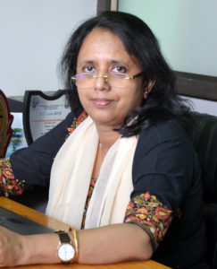
MIPS Seminar: Investigating and Imaging key molecular switches associated with Acquirement of Platinum-Taxol resistance in Epithelial Ovarian Cancer
Pritha Ray, Ph.D.
Principal Investigator & Scientific Officer F
Imaging Cell Signaling & Therapeutics Lab
ACTREC, Tata Memorial Center
Navi Mumbai, India
MIPS Seminar Series: Translational Opportunities in Glycoscience
Carolyn Bertozzi, PhD
Director, ChEM-H
Anne T. and Robert M. Bass Professor in the School of Humanities and Sciences
Professor, by courtesy, of Chemical and Systems Biology
Stanford University
Location: Zoom
Webinar URL: . https://stanford.zoom.us/j/94010708043
Dial: US: +1 650 724 9799 or +1 833 302 1536 (Toll Free)
Webinar ID: 940 1070 8043
Passcode: 659236
12:00pm – 12:45pm Seminar & Discussion
RSVP Here
ABSTRACT
Cell surface glycans constitute a rich biomolecular dataset that drives both normal and pathological processes. Their “readers” are glycan-binding receptors that can engage in cell-cell interactions and cell signaling. Our research focuses on mechanistic studies of glycan/receptor biology and applications of this knowledge to new therapeutic strategies. Our recent efforts center on pathogenic glycans in the tumor microenvironment and new therapeutic modalities based on the concept of targeted degradation.
ABOUT
Carolyn Bertozzi is the Baker Family Director of Stanford ChEM-H and the Anne T. and Robert M. Bass Professor of Humanities and Sciences in the Department of Chemistry at Stanford University. She is also an Investigator of the Howard Hughes Medical Institute. Her research focuses on profiling changes in cell surface glycosylation associated with cancer, inflammation and infection, and exploiting this information for development of diagnostic and therapeutic approaches, most recently in the area of immuno-oncology. She is an elected member of the National Academy of Medicine, the National Academy of Sciences, and the American Academy of Arts and Sciences. She also has been awarded the Lemelson-MIT Prize, a MacArthur Foundation Fellowship, the Chemistry for the Future Solvay Prize, among many others.
Hosted by: Katherine Ferrara, PhD
Sponsored by: Molecular Imaging Program at Stanford & the Department of Radiology
MIPS Seminar Series: “Convergent, translational research to improve human health”
Joseph M. DeSimone, PhD
Sanjiv Sam Gambhir Professor of Translational Medicine and Chemical Engineering
Departments of Radiology and Chemical Engineering
Graduate School of Business (by Courtesy)
Stanford University
Location: Zoom
Webinar URL: https://stanford.zoom.us/s/98460805010
Dial: +1 650 724 9799 or +1 833 302 1536
Webinar ID: 984 6080 5010
Passcode: 809226
12:00pm – 12:45pm Seminar & Discussion
RSVP Here
ABSTRACT
In many ways, manufacturing processes define what’s possible in society. Central to our interests in the DeSimone laboratory are opportunities to make things using cutting-edge fabrication technologies that can improve human health. This lecture will describe advances in nano- / micro-fabrication and 3D printing technologies that we have made and employed toward this end. Using novel perfluoropolyether materials synthesized in our lab in 2004, we invented the Particle Replication in Non-wetting Templates (PRINT) technology, a high-resolution imprint lithography-based process to fabricate nano- and micro-particles with precise and independent control over particle parameters (e.g. size, shape, modulus, composition, charge, surface chemistry). PRINT brought the precision and uniformity associated with computer industry manufacturing technologies to medicine, resulting in the launch of Liquidia Technologies (NASDAQ: LQDA) and opening new research paths, including to elucidate the influence of specific particle parameters in biological systems (Proc. Natl. Acad. Sci. USA 2008), and to reveal insights to inform the design of vaccines (J. Control. Release 2018), targeted therapeutics (Nano Letters 2015), and even synthetic blood (PNAS 2011). In 2015, we reported the invention of the Continuous Liquid Interface Production (CLIP) 3D printing technology (Science 2015), which overcame major fundamental limitations in polymer 3D printing—slowness, a very limited range of materials, and an inability to create parts with the mechanical and thermal properties needed for widespread, durable utility. By rethinking the physics and chemistry of 3D printing, we created CLIP to eliminate layer-by-layer fabrication altogether. A rapid, continuous process, CLIP generates production-grade parts and is now transforming how products are manufactured in industries including automotive, footwear, and medicine. For example, to help address shortages, CLIP recently enabled a new nasopharyngeal swab for COVID-19 diagnostic testing to go from concept to market in just 20 days, followed by a 400-patient clinical trial at Stanford. Academic laboratories are also using CLIP to pursue new medical device possibilities, including geometrically complex IVRs to optimize drug delivery and implantable chemotherapy absorbers to limit toxic side effects. Vast opportunities exist to use CLIP to pursue next-generation medical devices and prostheses. Moreover, CLIP can improve current approaches; for example, the fabrication of an iontophoretic device we invented several years ago (Sci. Transl. Med. 2015) to drive chemotherapeutics directly into hard-to-reach solid tumors is now being optimized for clinical trials with CLIP. New design opportunities also exist in early detection, for example to improve specimen collection, device performance (e.g. microfluidics, cell sorting, supporting growth and studies with human organoids), and imaging (e.g. PET detectors, ultrasound transducers). Here at Stanford, we are pursuing new 3D printing advances, including software treatment planning for digital therapeutic devices in pediatric medicine, as well as the design of a high-resolution printer capable of single-digit micron resolution to advance microneedle designs as a potent delivery platform for vaccines. The impact of our work on human health ultimately relies on our ability to enable a convergent research program to take shape that allows for new connections to be made among traditionally disparate disciplines and concepts, and to ensure that we maintain a consistent focus on the translational potential of our discoveries and advances.
ABOUT
Joseph M. DeSimone is the Sanjiv Sam Gambhir Professor of Translational Medicine and Chemical Engineering at Stanford University. He holds appointments in the Departments of Radiology and Chemical Engineering with a courtesy appointment in Stanford’s Graduate School of Business. Previously, DeSimone was a professor of chemistry at the University of North Carolina at Chapel Hill and of chemical engineering at North Carolina State University. He is also Co-founder, Board Chair, and former CEO (2014 – 2019) of the additive manufacturing company, Carbon.
DeSimone is responsible for numerous breakthroughs in his career in areas including green chemistry, medical devices, nanomedicine, and 3D printing, also co-founding several companies based on his research. He has published over 350 scientific articles and is a named inventor on over 200 issued patents. Additionally, he has mentored 80 students through Ph.D. completion in his career, half of whom are women and members of underrepresented groups in STEM. In 2016 DeSimone was recognized by President Barack Obama with the National Medal of Technology and Innovation, the highest U.S. honor for achievement and leadership in advancing technological progress. He is also one of only 25 individuals elected to all three branches of the U.S. National Academies (Sciences, Medicine, Engineering). DeSimone received his B.S. in Chemistry in 1986 from Ursinus College and his Ph.D. in Chemistry in 1990 from Virginia Tech.
Hosted by: Katherine Ferrara, PhD
Sponsored by: Molecular Imaging Program at Stanford & the Department of Radiology