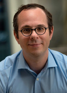
CEDSS: “The Origins and Detection of Lethal Prostate Cancer”
Paul Boutros, Ph.D., M.B.A.
Director, Cancer Data Sciences
UCLA
Please see zoom details below:
Meeting URL: https://stanford.zoom.us/s/93515779500
Dial: +1 650 724 9799 or +1 833 302 1536
Meeting ID: 935 1577 9500
Meeting Passcode: 767148
ABOUT
Boutros earned his B.Sc. degree from the University of Waterloo in Chemistry in 2004, and his Ph.D. degree from the University of Toronto, Canada, in Medical Biophysics in 2008. At Toronto, he also earned an executive M.B.A. from the Rothman School of Management. In 2008, Boutros started his independent research career at the Ontario Institute for Cancer Research first as a fellow (2008–2010) and then as principal investigator (2010–2018). He moved to California to join the UCLA faculty in 2018.
Hosted by: Utkan Demirci, Ph.D.
Sponsored by the Canary Center & the Department of Radiology
Stanford University – School of Medicine

ZOOM LINK HERE
“High Resolution Breast Diffusion Weighted Imaging”
Jessica McKay, PhD
ABSTRACT: Diffusion-weighted imaging (DWI) is a quantitative MRI method that measures the apparent diffusion coefficient (ADC) of water molecules, which reflects cell density and serves as an indication of malignancy. Unfortunately, however, the clinical value of DWI is severely limited by the undesirable features in images that common clinical methods produce, including large geometric distortions, ghosting and chemical shift artifacts, and insufficient spatial resolution. Thus, in order to exploit information encoded in diffusion characteristics and fully assess the clinical value of ADC measurements, it is first imperative to achieve technical advancements of DWI.
In this talk, I will largely focus on the background of breast DWI, providing the clinical motivation for this work and explaining the current standard in breast DWI and alternatives proposed throughout the literature. I will also present my PhD dissertation work in which a novel strategy for high resolution breast DWI was developed. The purpose of this work is to improve DWI methods for breast imaging at 3 Tesla to robustly provide diffusion-weighted images and ADC maps with anatomical quality and resolution. This project has two major parts: Nyquist ghost correction and the use of simultaneous multislice imaging (SMS) to achieve high resolution. Exploratory work was completed to characterize the Nyquist ghost in breast DWI, showing that, although the ghost is mostly linear, the three-line navigator is unreliable, especially in the presence of fat. A novel referenceless ghost correction, Ghost/Object minimization was developed that reduced the ghost in standard SE-EPI and advanced SMS. An advanced SMS method with axial reformatting (AR) is presented for high resolution breast DWI. In a reader study, AR-SMS was preferred by three breast radiologists compared to the standard SE-EPI and readout-segmented-EPI.
“Machine-learning Approach to Differentiation of Benign and Malignant Peripheral Nerve Sheath Tumors: A Multicenter Study”
Michael Zhang, MD
ABSTRACT: Clinicoradiologic differentiation between benign and malignant peripheral nerve sheath tumors (PNSTs) is a diagnostic challenge with important management implications. We sought to develop a radiomics classifier based on 900 features extracted from gadolinium-enhanced, T1-weighted MRI, using the Quantitative Imaging Feature Pipeline and the PyRadiomics package. Additional patient-specific clinical variables were recorded. A radiomic signature was derived from least absolute shrinkage and selection operator, followed by gradient boost machine learning. A training and test set were selected randomly in a 70:30 ratio. We further evaluated the performance of radiomics-based classifier models against human readers of varying medical-training backgrounds. Following image pre-processing, 95 malignant and 171 benign PNSTs were available. The final classifier included 21 features and achieved a sensitivity 0.676, specificity 0.882, and area under the curve (AUC) 0.845. Collectively, human readers achieved sensitivity 0.684, specificity 0.742, and AUC 0.704. We concluded that radiomics using routine gadolinium enhanced, T1-weighted MRI sequences and clinical features can aid in the evaluation of PNSTs, particularly by increasing specificity for diagnosing malignancy. Further improvement may be achieved with incorporation of additional imaging sequences.
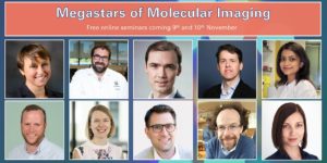
About this Event
This two-day seminar series brings together the brightest minds in molecular imaging to discuss their latest research. Each talk will last 30 min with a full 15 min dedicated for Q&A, related both to their science and their career. So bring that question you’ve always wanted to ask!
Agenda (Note all times are in GMT)
Day 1 (9th November)
13:30 – Ferdia Gallagher (Cambridge University) ‘Clinical imaging of tumour metabolism’
14:15 – David Lewis (CRUK Beatson Institute) ‘Illuminating metabolic vulnerabilities in lung cancer’
15:00 – Federica Pisaneschi (MD Anderson Cancer Center) ‘Imaging ROS burst during myeloid cell activation with 4-[18F]fluoronaphthol’
15:45 – Break
16:00 – Adam Shuhendler (University of Ottawa) ‘Activity-based Sensing by CEST-MRI’
16:45 – Israt Alam (Stanford University) ‘A tale of two biomarkers: visualizing T cell activation with immunoPET’
Day 2 (10th November)
13:30 – André Neves (Cambridge University) ‘Molecular imaging of aberrant glycosylation in cancer’
14:15 – Gilbert Fruhwirth (King’s College London) ‘How non-invasive in vivo cell tracking supports the development of advanced immunotherapeutics’
15:00 – Sarah Bohndiek (Cambridge University) ‘Shedding light on the tumour vasculature’
15:45 – Break
16:00 – John Ronald (Robarts Research Institute) ‘Reporter genes and genome editing for MRI cell tracking: Maybe MRI doesn’t suck?’
16:45 – Michelle James (Stanford University) ‘Shedding light on the immune system to improve detection and treatment of brain diseases using PET’

Ge Wang, PhD
Clark & Crossan Endowed Chair Professor
Director of the Biomedical Imaging Center
Rensselaer Polytechnic Institute
Troy, New York
Abstract:
AI-based tomography is an important application and a new frontier of machine learning. AI, especially deep learning, has been widely used in computer vision and image analysis, which deal with existing images, improve them, and produce features. Since 2016, deep learning techniques are actively researched for tomography in the context of medicine. Tomographic reconstruction produces images of multi-dimensional structures from externally measured “encoded” data in the form of various transforms (integrals, harmonics, and so on). In this presentation, we provide a general background, highlight representative results, and discuss key issues that need to be addressed in this emerging field.
About:
AI-based X-ray Imaging System (AXIS) lab is led by Dr. Ge Wang, affiliated with the Department of Biomedical Engineering at Rensselaer Polytechnic Institute and the Center for Biotechnology and Interdisciplinary Studies in the Biomedical Imaging Center. AXIS lab focuses on innovation and translation of x-ray computed tomography, optical molecular tomography, multi-scale and multi-modality imaging, and AI/machine learning for image reconstruction and analysis, and has been continuously well funded by federal agencies and leading companies. AXIS group collaborates with Stanford, Harvard, Cornell, MSK, UTSW, Yale, GE, Hologic, and others, to develop theories, methods, software, systems, applications, and workflows.

CEDSS: Systematic identification of fluid-based biomarkers for ovarian and prostate cancer
Thomas Kislinger, Ph.D.
Professor & Chair
Department of Medical Biophysics
University of Toronto
Senior Scientist
Princess Margaret Cancer Centre
Zoom Webinar Details
Meeting URL: https://stanford.zoom.us/s/94878578384
Dial: +1 650 724 9799 or +1 833 302 1536
Webinar ID: 948 7857 8384
Passcode: 692692
Register Here
ABOUT
Thomas Kislinger received his MSc in Analytical Chemistry from the University of Munich, Germany (1998). He completed his PhD in 2001, investigating the role of Advanced Glycation Endproducts in diabetic vascular complications at the University of Erlangen, Germany and Columbia University, New York. Between 2002 and 2006 he completed a post-doctoral fellowship at the University of Toronto. In 2006 he joined the Princess Margaret Cancer Centre as an independent investigator. Dr. Kislinger holds positions as Senior Scientist at the Princess Margaret Cancer Centre and as Professor and Chair at the University of Toronto in the Department of Medical Biophysics. The Kislinger lab applies proteomics technologies to translational and basic cancer biology. This includes the development of novel proteomics methodologies, identification of liquid biopsy signatures and the molecular identification of novel cell surface markers.
Hosted by: Utkan Demirci, Ph.D.
Sponsored by: The Canary Center & the Department of Radiology
Stanford University – School of Medicine
Intersection of Imaging and Therapeutics – MIPS Mini-Retreat
Hosted by: Dr. Heike Daldrup-Link, MD & Dr. Katherine Ferrara, PhD
Sponsored by: Department of Radiology, Molecular Imaging Program at Stanford
The MIPS Mini-retreat series brings together members of the MIPS and greater School of Medicine community to discuss current opportunities for research and collaborations. Each month we will discuss a different topic and we invite all those interested to attend. The mini-retreats will be roughly 1.5-2 hours in length with ample time for discussion throughout. We hope you can join us and spark new collaborations!
Zoom Webinar Information
Webinar URL: https://stanford.zoom.us/j/96227145646
Dial: US: +1 650 724 9799 or +1 833 302 1536 (Toll Free)
Webinar ID: 962 2714 5646
Passcode: 3039816
Agenda (all times are in PST)
8:00-8:05 AM – Opening Remarks – Katherine Ferrara, PhD
8:05-8:20 AM – Image-guided Therapy – Heike Daldrup-Link, MD
8:20-8:35 AM – Imaging Antibody Distribution – Eben Rosenthal, MD
8:35-8:50 AM – MRgFUS and Pancreatic Cancer – Pejman Ghanouni, MD, PhD
8:50-9:05 AM – RefleXion – Lucas Kas Vitzhum, MD
9:05-10:00 AM – Discussion – Moderated by: Katherine Ferrara, PhD
MIPS Seminar Series: Translational Opportunities in Glycoscience
Carolyn Bertozzi, PhD
Director, ChEM-H
Anne T. and Robert M. Bass Professor in the School of Humanities and Sciences
Professor, by courtesy, of Chemical and Systems Biology
Stanford University
Location: Zoom
Webinar URL: . https://stanford.zoom.us/j/94010708043
Dial: US: +1 650 724 9799 or +1 833 302 1536 (Toll Free)
Webinar ID: 940 1070 8043
Passcode: 659236
12:00pm – 12:45pm Seminar & Discussion
RSVP Here
ABSTRACT
Cell surface glycans constitute a rich biomolecular dataset that drives both normal and pathological processes. Their “readers” are glycan-binding receptors that can engage in cell-cell interactions and cell signaling. Our research focuses on mechanistic studies of glycan/receptor biology and applications of this knowledge to new therapeutic strategies. Our recent efforts center on pathogenic glycans in the tumor microenvironment and new therapeutic modalities based on the concept of targeted degradation.
ABOUT
Carolyn Bertozzi is the Baker Family Director of Stanford ChEM-H and the Anne T. and Robert M. Bass Professor of Humanities and Sciences in the Department of Chemistry at Stanford University. She is also an Investigator of the Howard Hughes Medical Institute. Her research focuses on profiling changes in cell surface glycosylation associated with cancer, inflammation and infection, and exploiting this information for development of diagnostic and therapeutic approaches, most recently in the area of immuno-oncology. She is an elected member of the National Academy of Medicine, the National Academy of Sciences, and the American Academy of Arts and Sciences. She also has been awarded the Lemelson-MIT Prize, a MacArthur Foundation Fellowship, the Chemistry for the Future Solvay Prize, among many others.
Hosted by: Katherine Ferrara, PhD
Sponsored by: Molecular Imaging Program at Stanford & the Department of Radiology

Imaging Immune Cell Modulation – MIPS Mini-Retreat
Hosted by: Dr. Katherine Ferrara, PhD
Sponsored by: Department of Radiology, Molecular Imaging Program at Stanford
The MIPS Mini-retreat series brings together members of the MIPS and greater School of Medicine community to discuss current opportunities for research and collaborations. Each month we will discuss a different topic and we invite all those interested to attend. The mini-retreats will be roughly 1.5-2 hours in length with ample time for discussion throughout. We hope you can join us and spark new collaborations!
Zoom Webinar Information
Webinar URL: https://stanford.zoom.us/j/94969801773
Dial: US: +1 650 724 9799 or +1 833 302 1536 (Toll Free)
Webinar ID: 949 6980 1773
Passcode: 292995
Agenda (all times are in PST)
8:00-8:05 AM – Opening Remarks – Katherine Ferrara, PhD
8:05-8:25 AM – Imaging CAR-T cells – Clinical Need – Crystal Mackall, MD
8:25-8:45 AM – Imaging Immune Cells in Cancer – Clinical Need – Ronald Levy, MD
8:45-9:05 AM – Imaging Approaches – Anna Wu, PhD
9:05-10:00 AM – Discussion – Moderated by: Katherine Ferrara, PhD
MIPS Seminar Series: “Convergent, translational research to improve human health”
Joseph M. DeSimone, PhD
Sanjiv Sam Gambhir Professor of Translational Medicine and Chemical Engineering
Departments of Radiology and Chemical Engineering
Graduate School of Business (by Courtesy)
Stanford University
Location: Zoom
Webinar URL: https://stanford.zoom.us/s/98460805010
Dial: +1 650 724 9799 or +1 833 302 1536
Webinar ID: 984 6080 5010
Passcode: 809226
12:00pm – 12:45pm Seminar & Discussion
RSVP Here
ABSTRACT
In many ways, manufacturing processes define what’s possible in society. Central to our interests in the DeSimone laboratory are opportunities to make things using cutting-edge fabrication technologies that can improve human health. This lecture will describe advances in nano- / micro-fabrication and 3D printing technologies that we have made and employed toward this end. Using novel perfluoropolyether materials synthesized in our lab in 2004, we invented the Particle Replication in Non-wetting Templates (PRINT) technology, a high-resolution imprint lithography-based process to fabricate nano- and micro-particles with precise and independent control over particle parameters (e.g. size, shape, modulus, composition, charge, surface chemistry). PRINT brought the precision and uniformity associated with computer industry manufacturing technologies to medicine, resulting in the launch of Liquidia Technologies (NASDAQ: LQDA) and opening new research paths, including to elucidate the influence of specific particle parameters in biological systems (Proc. Natl. Acad. Sci. USA 2008), and to reveal insights to inform the design of vaccines (J. Control. Release 2018), targeted therapeutics (Nano Letters 2015), and even synthetic blood (PNAS 2011). In 2015, we reported the invention of the Continuous Liquid Interface Production (CLIP) 3D printing technology (Science 2015), which overcame major fundamental limitations in polymer 3D printing—slowness, a very limited range of materials, and an inability to create parts with the mechanical and thermal properties needed for widespread, durable utility. By rethinking the physics and chemistry of 3D printing, we created CLIP to eliminate layer-by-layer fabrication altogether. A rapid, continuous process, CLIP generates production-grade parts and is now transforming how products are manufactured in industries including automotive, footwear, and medicine. For example, to help address shortages, CLIP recently enabled a new nasopharyngeal swab for COVID-19 diagnostic testing to go from concept to market in just 20 days, followed by a 400-patient clinical trial at Stanford. Academic laboratories are also using CLIP to pursue new medical device possibilities, including geometrically complex IVRs to optimize drug delivery and implantable chemotherapy absorbers to limit toxic side effects. Vast opportunities exist to use CLIP to pursue next-generation medical devices and prostheses. Moreover, CLIP can improve current approaches; for example, the fabrication of an iontophoretic device we invented several years ago (Sci. Transl. Med. 2015) to drive chemotherapeutics directly into hard-to-reach solid tumors is now being optimized for clinical trials with CLIP. New design opportunities also exist in early detection, for example to improve specimen collection, device performance (e.g. microfluidics, cell sorting, supporting growth and studies with human organoids), and imaging (e.g. PET detectors, ultrasound transducers). Here at Stanford, we are pursuing new 3D printing advances, including software treatment planning for digital therapeutic devices in pediatric medicine, as well as the design of a high-resolution printer capable of single-digit micron resolution to advance microneedle designs as a potent delivery platform for vaccines. The impact of our work on human health ultimately relies on our ability to enable a convergent research program to take shape that allows for new connections to be made among traditionally disparate disciplines and concepts, and to ensure that we maintain a consistent focus on the translational potential of our discoveries and advances.
ABOUT
Joseph M. DeSimone is the Sanjiv Sam Gambhir Professor of Translational Medicine and Chemical Engineering at Stanford University. He holds appointments in the Departments of Radiology and Chemical Engineering with a courtesy appointment in Stanford’s Graduate School of Business. Previously, DeSimone was a professor of chemistry at the University of North Carolina at Chapel Hill and of chemical engineering at North Carolina State University. He is also Co-founder, Board Chair, and former CEO (2014 – 2019) of the additive manufacturing company, Carbon.
DeSimone is responsible for numerous breakthroughs in his career in areas including green chemistry, medical devices, nanomedicine, and 3D printing, also co-founding several companies based on his research. He has published over 350 scientific articles and is a named inventor on over 200 issued patents. Additionally, he has mentored 80 students through Ph.D. completion in his career, half of whom are women and members of underrepresented groups in STEM. In 2016 DeSimone was recognized by President Barack Obama with the National Medal of Technology and Innovation, the highest U.S. honor for achievement and leadership in advancing technological progress. He is also one of only 25 individuals elected to all three branches of the U.S. National Academies (Sciences, Medicine, Engineering). DeSimone received his B.S. in Chemistry in 1986 from Ursinus College and his Ph.D. in Chemistry in 1990 from Virginia Tech.
Hosted by: Katherine Ferrara, PhD
Sponsored by: Molecular Imaging Program at Stanford & the Department of Radiology

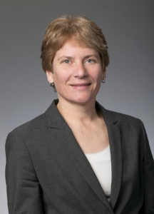
CEDSS: Disseminated cell hybrids as biomarkers for cancer detection, prognosis and treatment response
Melissa Wong, Ph.D.
Associate Professor and Vice Chair
Department of Cell, Development and Cancer Biology
Program Co-Lead, Knight Cancer Institute
Oregon Health & Science University
Zoom Details
Meeting URL: https://stanford.zoom.us/s/98184098662
Dial: US: +1 650 724 9799 or +1 833 302 1536 (Toll Free)
Meeting ID: 981 8409 8662
Passcode: 084321
ABSTRACT
Metastatic progression defines the final stages of tumor evolution and underlies the majority of cancer-related deaths. The heterogeneity in disseminated tumor cell populations capable of seeding and growing in distant organ sites contributes to the development of treatment resistant disease. We recently reported the identification of a novel tumor-derived cell population, circulating hybrid cells (CHCs), harboring attributes from both macrophages and neoplastic cells, including functional characteristics important to metastatic spread. These disseminated hybrids outnumber conventionally defined circulating tumor cells (CTCs) in cancer patients. It is unknown if CHCs represent a generalized cancer mechanism for cell dissemination, or if this population is relevant to the metastatic cascade. We detect CHCs in the peripheral blood of patients with cancer in myriad disease sites encompassing epithelial and non-epithelial malignancies. Further, we demonstrate that in vivo-derived hybrid cells harbor tumor-initiating capacity in murine cancer models and that CHCs from human breast cancer patients express stem cell antigens, features consistent with the ability to seed and grow at metastatic sites. We reveal heterogeneity of CHC phenotypes reflect key tumor features, including oncogenic mutations and functional protein expression. Importantly, this novel population of disseminated neoplastic cells opens a new area in cancer biology and renewed opportunity for battling metastatic disease.
ABOUT
The research focus of the Wong laboratory revolves around understanding the regulatory mechanisms that control epithelial stem cell homeostasis and their expansion in developmental, homeostasis and disease contexts, including cancer. I have substantial training and experience in intestinal stem cell investigation leveraging in vivo and ex vivo modeling, as well as in myriad cutting edge technologies (i.e. cyCIF, scRNA-seq). My publication record spans my post-doctoral fellowship in Dr. Jeffrey Gordon’s laboratory at Washington University School of Medicine, to studies in my own laboratory at Oregon Health & Science University. Our research impacts the understanding of regulatory mechanisms that govern cell state in the context of the evolving tissue microenvironment and changing cell signaling landscape, in development and disease.
Our studies in stem cell regulation led to the intriguing finding that stem cells can fuse with tissue macrophages in the context of injury repair and may impact tissue regeneration. We have extended these findings to the cancer setting, where cancer-macrophage fusions are detectible in primary and metastatic tumors, and my group recently identified and characterized these cells as a novel circulating tumor cell population. Importantly, our studies in cell culture, in mice and humans provide an indepth evaluation of hybrid cells to set the foundation for continued investigations into their biology, impact on disease progression or tissue regeneration, and use as a biomarker for disease burden. Importantly, we coined the term, circulating hybrid cell (CHC) for this novel population and reported they exist at higher levels than conventionally defined circulating tumor cells in the peripheral blood of cancer patients. This work was published in 2018 and highlighted by Science Magazine as one of the top ten publications in the cancer field in the science family journals. The science proposed in this U01 application leverage hybrid cell biology to assess treatment response and resistance in breast cancer patients undergoing targeted therapy. Our proposal leverages active collaborations with Dr. Young Hwan Young’s group to synergize biology with computation, as well as a number of other valuable collaborators to ensure success of the proposed, cutting-edge science.
Hosted by: Utkan Demirci, Ph.D.
Sponsored by: The Canary Center & the Department of Radiology
Stanford University – School of Medicine