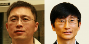
Thursday MIPS Roundtable: Faculty Lab Showcase
1:30-2:00 PM | Dr. Jianghong Rao, Ph.D.
Cellular and Molecular Imaging Laboratory (CMIL)
Professor of Radiology and, by courtesy, of Chemistry
Stanford University
2:00-2:30 PM | Dr. Zhen Cheng, Ph.D.
Cancer Molecular Imaging Chemistry Laboratory (CMICL)
Associate Professor of Radiology
Stanford University
MIPS Roundtables will be every Thursday from 1:30-2:30pm showcasing various topics and are open to all interested.
Please note Zoom information does change week to week.
5/28 Meeting URL: https://stanford.zoom.us/j/92834097988
Dial: +1 650 724 9799 or +1 833 302 1536
Meeting ID: 928 3409 7988

Thursday MIPS Roundtable: Meet our MIPS Instructors
1:30-2:00 PM | Dr. Katie Wilson, Ph.D.
“Optical and Acoustic Molecular Imaging to Identify Lymph Node Metastasis in Head and Neck Cancer.”
Instructor, Radiology
Stanford University
2:00-2:30 PM | Dr. Corinne Beinat, Ph.D.
“Molecular Imaging of Tumor Metabolism”
Instructor, Radiology
Stanford University
MIPS Roundtables are every other Thursday from 1:30-2:30pm showcasing various topics and are open to all interested.
Please note Zoom information does change week to week.
6/11 Meeting URL: https://stanford.zoom.us/j/95475611159
Dial: +1 650 724 9799 or +1 833 302 1536
Meeting ID: 954 7561 1159

Thursday MIPS Roundtable: Faculty Lab Showcase
MIPS Roundtables are every other Thursday from 1:30-2:30pm showcasing various topics and are open to all interested.
1:30-2:00 PM | Dr. Brian Rutt, Ph.D.
Cellular & Molecular MRI Laboratory (CMMRIL)
Professor of Radiology
Stanford University
2:00-2:30 PM | Dr. Kathy Ferrara, Ph.D.
Ferrara Laboratory: Image-guided Drug Delivery
Professor of Radiology
Stanford University
Please note Zoom information does change week to week.
6/25 Webinar URL: https://stanford.zoom.us/j/91635637393?pwd=c09vUXYyeU5VeHJBaUJVRHQrT3FJdz09
Dial: +1 650 724 9799 or +1 833 302 1536
Webinar ID: 916 3563 7393
Webinar Password: 271364
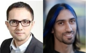
Thursday MIPS Roundtable: Meet our MIPS Instructors
MIPS Roundtables are every other Thursday from 1:30-2:30pm showcasing various topics and are open to all interested. Note we will take a break through late July and August.
1:30-2:00 PM | Dr. Ahmed El Kaffas, Ph.D.
Translational Ultrasound for Tissue Characterization and Stimulation
Instructor, Radiology
Stanford University
2:00-2:30 PM | Dr. Brett Fite, Ph.D.
Combining Focal and Immunotherapies
Instructor, Radiology
Stanford University
Please note Zoom information does change week to week.
7/9 Webinar URL: https://stanford.zoom.us/j/91909413178
Dial: +1 650 724 9799 or +1 833 302 1536
Webinar ID: 919 0941 3178
Password: 572746

Thursday MIPS Roundtable: Meet our MIPS Instructors
MIPS Roundtables are Thursdays from 1:30-2:30pm showcasing various topics and are open to all interested. Note this will be our last summer Roundtable and we will take a break through late July and August.
1:30-2:00 PM | Dr. Josquin Foiret, Ph.D.
High throughput ultrasound imaging for improved diagnosis
Instructor, Radiology
Stanford University
2:00-2:30 PM | Dr. Jinghang Xie, Ph.D.
TESLA probes for imaging T cell-mediated cytotoxic response to immunotherapy
Instructor, Radiology
Stanford University
Please note Zoom information does change week to week.
7/16 Webinar URL: https://stanford.zoom.us/j/94952044130
Dial: +1 650 724 9799 or +1 833 302 1536
Webinar ID: 949 5204 4130
Password: 963699
Stanford Molecular Imaging Scholars (SMIS) Program Quarterly Seminar
Zoom meeting: https://stanford.zoom.us/j/99117388314?pwd=R29OSjlTdUt0a3pLaG5Zc1BFNTJIUT09
Password: 922183
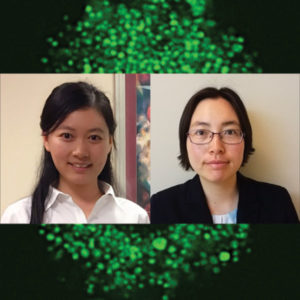
Guolan Lu, PhD
Mentor: Eben Rosenthal, MD; Garry Nolan, PhD
“Co-administered Antibody Improves the Penetration of Antibody-Dye Conjugates into Human Cancers: Implications for AntibodyDrug Conjugates”
Dianna Jeong, PhD
Mentors: Craig Levin, PhD; Shan Wang, PhD
“Novel Detection Approaches for Achieving Ultra-fast time resolution for PET”

ZOOM LINK HERE
“High Resolution Breast Diffusion Weighted Imaging”
Jessica McKay, PhD
ABSTRACT: Diffusion-weighted imaging (DWI) is a quantitative MRI method that measures the apparent diffusion coefficient (ADC) of water molecules, which reflects cell density and serves as an indication of malignancy. Unfortunately, however, the clinical value of DWI is severely limited by the undesirable features in images that common clinical methods produce, including large geometric distortions, ghosting and chemical shift artifacts, and insufficient spatial resolution. Thus, in order to exploit information encoded in diffusion characteristics and fully assess the clinical value of ADC measurements, it is first imperative to achieve technical advancements of DWI.
In this talk, I will largely focus on the background of breast DWI, providing the clinical motivation for this work and explaining the current standard in breast DWI and alternatives proposed throughout the literature. I will also present my PhD dissertation work in which a novel strategy for high resolution breast DWI was developed. The purpose of this work is to improve DWI methods for breast imaging at 3 Tesla to robustly provide diffusion-weighted images and ADC maps with anatomical quality and resolution. This project has two major parts: Nyquist ghost correction and the use of simultaneous multislice imaging (SMS) to achieve high resolution. Exploratory work was completed to characterize the Nyquist ghost in breast DWI, showing that, although the ghost is mostly linear, the three-line navigator is unreliable, especially in the presence of fat. A novel referenceless ghost correction, Ghost/Object minimization was developed that reduced the ghost in standard SE-EPI and advanced SMS. An advanced SMS method with axial reformatting (AR) is presented for high resolution breast DWI. In a reader study, AR-SMS was preferred by three breast radiologists compared to the standard SE-EPI and readout-segmented-EPI.
“Machine-learning Approach to Differentiation of Benign and Malignant Peripheral Nerve Sheath Tumors: A Multicenter Study”
Michael Zhang, MD
ABSTRACT: Clinicoradiologic differentiation between benign and malignant peripheral nerve sheath tumors (PNSTs) is a diagnostic challenge with important management implications. We sought to develop a radiomics classifier based on 900 features extracted from gadolinium-enhanced, T1-weighted MRI, using the Quantitative Imaging Feature Pipeline and the PyRadiomics package. Additional patient-specific clinical variables were recorded. A radiomic signature was derived from least absolute shrinkage and selection operator, followed by gradient boost machine learning. A training and test set were selected randomly in a 70:30 ratio. We further evaluated the performance of radiomics-based classifier models against human readers of varying medical-training backgrounds. Following image pre-processing, 95 malignant and 171 benign PNSTs were available. The final classifier included 21 features and achieved a sensitivity 0.676, specificity 0.882, and area under the curve (AUC) 0.845. Collectively, human readers achieved sensitivity 0.684, specificity 0.742, and AUC 0.704. We concluded that radiomics using routine gadolinium enhanced, T1-weighted MRI sequences and clinical features can aid in the evaluation of PNSTs, particularly by increasing specificity for diagnosing malignancy. Further improvement may be achieved with incorporation of additional imaging sequences.

About this Event
This two-day seminar series brings together the brightest minds in molecular imaging to discuss their latest research. Each talk will last 30 min with a full 15 min dedicated for Q&A, related both to their science and their career. So bring that question you’ve always wanted to ask!
Agenda (Note all times are in GMT)
Day 1 (9th November)
13:30 – Ferdia Gallagher (Cambridge University) ‘Clinical imaging of tumour metabolism’
14:15 – David Lewis (CRUK Beatson Institute) ‘Illuminating metabolic vulnerabilities in lung cancer’
15:00 – Federica Pisaneschi (MD Anderson Cancer Center) ‘Imaging ROS burst during myeloid cell activation with 4-[18F]fluoronaphthol’
15:45 – Break
16:00 – Adam Shuhendler (University of Ottawa) ‘Activity-based Sensing by CEST-MRI’
16:45 – Israt Alam (Stanford University) ‘A tale of two biomarkers: visualizing T cell activation with immunoPET’
Day 2 (10th November)
13:30 – André Neves (Cambridge University) ‘Molecular imaging of aberrant glycosylation in cancer’
14:15 – Gilbert Fruhwirth (King’s College London) ‘How non-invasive in vivo cell tracking supports the development of advanced immunotherapeutics’
15:00 – Sarah Bohndiek (Cambridge University) ‘Shedding light on the tumour vasculature’
15:45 – Break
16:00 – John Ronald (Robarts Research Institute) ‘Reporter genes and genome editing for MRI cell tracking: Maybe MRI doesn’t suck?’
16:45 – Michelle James (Stanford University) ‘Shedding light on the immune system to improve detection and treatment of brain diseases using PET’

Ge Wang, PhD
Clark & Crossan Endowed Chair Professor
Director of the Biomedical Imaging Center
Rensselaer Polytechnic Institute
Troy, New York
Abstract:
AI-based tomography is an important application and a new frontier of machine learning. AI, especially deep learning, has been widely used in computer vision and image analysis, which deal with existing images, improve them, and produce features. Since 2016, deep learning techniques are actively researched for tomography in the context of medicine. Tomographic reconstruction produces images of multi-dimensional structures from externally measured “encoded” data in the form of various transforms (integrals, harmonics, and so on). In this presentation, we provide a general background, highlight representative results, and discuss key issues that need to be addressed in this emerging field.
About:
AI-based X-ray Imaging System (AXIS) lab is led by Dr. Ge Wang, affiliated with the Department of Biomedical Engineering at Rensselaer Polytechnic Institute and the Center for Biotechnology and Interdisciplinary Studies in the Biomedical Imaging Center. AXIS lab focuses on innovation and translation of x-ray computed tomography, optical molecular tomography, multi-scale and multi-modality imaging, and AI/machine learning for image reconstruction and analysis, and has been continuously well funded by federal agencies and leading companies. AXIS group collaborates with Stanford, Harvard, Cornell, MSK, UTSW, Yale, GE, Hologic, and others, to develop theories, methods, software, systems, applications, and workflows.
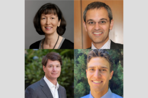
Intersection of Imaging and Therapeutics – MIPS Mini-Retreat
Hosted by: Dr. Heike Daldrup-Link, MD & Dr. Katherine Ferrara, PhD
Sponsored by: Department of Radiology, Molecular Imaging Program at Stanford
The MIPS Mini-retreat series brings together members of the MIPS and greater School of Medicine community to discuss current opportunities for research and collaborations. Each month we will discuss a different topic and we invite all those interested to attend. The mini-retreats will be roughly 1.5-2 hours in length with ample time for discussion throughout. We hope you can join us and spark new collaborations!
Zoom Webinar Information
Webinar URL: https://stanford.zoom.us/j/96227145646
Dial: US: +1 650 724 9799 or +1 833 302 1536 (Toll Free)
Webinar ID: 962 2714 5646
Passcode: 3039816
Agenda (all times are in PST)
8:00-8:05 AM – Opening Remarks – Katherine Ferrara, PhD
8:05-8:20 AM – Image-guided Therapy – Heike Daldrup-Link, MD
8:20-8:35 AM – Imaging Antibody Distribution – Eben Rosenthal, MD
8:35-8:50 AM – MRgFUS and Pancreatic Cancer – Pejman Ghanouni, MD, PhD
8:50-9:05 AM – RefleXion – Lucas Kas Vitzhum, MD
9:05-10:00 AM – Discussion – Moderated by: Katherine Ferrara, PhD