
PHIND Seminar Series: What is in your sweat and what can it mean for health and disease?
11:00 AM – 12:00 PM: Seminar & Discussion
RSVP: https://www.onlineregistrationcenter.com/VShankar
Presenter:
Vishnu Shankar, M.S.
Department of Chemistry
Stanford University
Principal Investigators:
Michael Snyder, Ph.D.
Stanford W. Ascherman, MD, FACS Professor in Genetics
Stanford University
Robert Tibshirani, Ph.D.
Professor of Biomedical Data Science and of Statistics
Stanford University
Richard Zare, Ph.D.
Marguerite Blake Wilbur Professor in Natural Science and Professor, by courtesy, of Physics
Stanford University
Location
Webinar URL: https://stanford.zoom.us/s/99817512229?pwd=QitCTjRXMEdBTWZyd29MTHYyNU5Xdz09
Dial: +1 650 724 9799 or +1 833 302 1536
Webinar ID: 998 1751 2229
Password: 489011
ABSTRACT
Sweat is a complex fluid known to be rich in electrolytes, small molecules, and fatty acids. Although adults can sweat up to 10 liters per day, little is still known about the chemical composition of sweat, how this changes, and what are its implications for health and disease. We demonstrate a powerful approach to help elucidate this link, where collecting samples simply requires swabbing a glass slide across one’s forehead in less than 30 seconds. Using the combination of desorption electrospray ionization mass spectrometry and statistical machine learning, our approach can successfully detect over 10,000 metabolites in sweat and identify metabolic changes in the sweat profile related to gender, age, and disease. As an example, we demonstrate in a cohort of 65 subjects the possibility of using just a few metabolites detected in sweat to successfully identify patients with renal disease. More generally, our approach suggests the possibility of using the sweat profile to non-invasively assess individual risk for metabolic diseases in the theme of “Precision Medicine.”
ABOUT VISHNU SHANKAR
Vishnu Shankar recently graduated with his master’s degree in computer science, with a specialization in artificial intelligence from Stanford University. He completed his bachelor’s degree with honors in mathematical and computational sciences in 2018, also at Stanford, with his senior thesis on Bayesian networks for incorporating effect modifiers in meta-analysis. In addition, his background spans biology, mathematics, chemistry, statistics, operations research, physics, and computing. Vishnu has published 6 papers and 3 articles in fields including protein structural prediction, comparison of clinical guidelines cost-effectiveness in type 2 diabetes, development of programs to combat mental illness, cancer diagnosis with analytical chemistry and machine learning, and related areas. He is also the founder of the CARES organization to support peer student wellness at college campuses, for which he won the Asoka Youth Changemaker award sponsored by Boehringer Ingelheim. Vishnu has enthusiastically pursued science since his middle school days and has worked on demonstrating the possibilities of DNA computing, simulating protein folding, studying the genetic modifications in fruits and vegetables, and more. He has been recognized for his scientific research as an Intel Science Talent finalist, Google Science Fair Regional finalist, recipient of the American Institute of Aeronautics and Astronautics Excellence award for his work on epidemiology modeling. Vishnu has interned at Caltech and Genentech, where he applied experimental techniques to purify and study protein behavior including dialysis, titration, chromatography for early stage drug development. He was also selected as one of the two high school students to represent the western US Confucius Institute in Student Leaders Exchange Program in China as a non-native Mandarin speaker.
Hosted by: Sanjiv Sam Gambhir, M.D., Ph.D.
Sponsored by the PHIND Center and the Department of Radiology
PHIND Seminar Series: Identifying Fibroblasts Subtypes Contributing to the Progression of Preinvasive to Invasive Lung Adenocarcinoma
Sylvia Plevritis, Ph.D.
Professor of Biomedical Data Science and of Radiology
Integrative Biomedical Imaging Informatics at Stanford
Stanford University
Location
Webinar URL: https://stanford.zoom.us/s/93945120934?pwd=a29GNjFCUzBtWjRsbFdnUnVUOTMzUT09
Dial: +1 650 724 9799 or +1 833 302 1536
Webinar ID: 939 4512 0934
Password: 767148
11:00am – 12:00pm Seminar & Discussion
RSVP: https://www.onlineregistrationcenter.com/SPlevritis
ABOUT
Dr. Sylvia K. Plevritis is Professor and Chair of Biomedical Data Science, Professor of Radiology at Stanford University and Program Director of the Stanford Biomedical Informatics Graduate Training Program. Dr. Plevritis leads a computational biology cancer research program that bridges genomics, imaging and population sciences to decipher properties of cancer progression and treatment response. Dr. Plevritis received her Ph.D. in Electrical Engineering and M.S. in Health Services Research, both from Stanford University, with a focus on cancer imaging physics and modeling cancer outcomes, respectively. She has had a primary authorship role on over 100 scientific cancer-related articles. She is a fellow of the American Institute for Medical and Biological Engineering (AIMBE) and Distinguished Investigator in the Academy of Radiology Research. She serves on the NCI Board of Scientific Advisors, the Program Leadership Committee of the Stanford Cancer Institute and the Leadership Council of the Stanford Bio-X Program. Dr. Plevritis has served on numerous NIH study sections, chaired scientific programs for the several professional societies including the American Association for Cancer Research (AACR) and presented keynote lectures across multiple scales of computational cancer biology. Currently, she is the Program Director of the Stanford Center in Cancer Systems Biology (CCSB) and has been a Principal Investigator with the NCI Cancer Intervention Surveillance Network (CISNET) for over fifteen years. She has served as Program Director of the Stanford Cancer Systems Biology Scholars Program (CSBS), and co-Division Chief of Integrative Biomedical Imaging Informatics at Stanford (IBIIS).
Sponsored by the PHIND Center and the Department of Radiology
PHIND Seminar Series: Maintaining Competitive Advantage through Intellectual Capital
Soody Tronson, M.S., J.D.
Founder
Presque
Location & Timing
11:00am – 12:00pm Seminar & Discussion
Webinar URL: https://stanford.zoom.us/s/98817009379?pwd=U21pYnpCTUlxY3U2TEh4RmhvMXpvQT09
Dial: +1 650 724 9799 or +1 833 302 1536
Webinar ID: 988 1700 9379
Password: 767148
RSVP: https://www.onlineregistrationcenter.com/STronson
ABSTRACT
Digital health has provided a range of solutions, including health and wellness-related management, machine learning algorithms for pattern recognition, AI for genetic analysis, AI-enhanced clinical decision making, and virtual doctors that use AI for patient intake triage. Intellectual property can play an important role in providing a competitive advantage in the digital health industry. We will explore opportunities and challenges for protecting digital health technologies during the program, including patent eligibility challenges and the use of trade secrets.
ABOUT SOODY TRONSON
Soody Tronson is Founding Managing Counsel at STLG Law Firm, counseling domestic and international clients in IP and technology transactions in a wide range of technologies. In 2016 she formed, Presque, a company developing a line of medical devices for mothers and infants.
Soody has over 25 years of operational experience in technology, business, management, and law in start-up and fortune 100 companies, including Schering Plough Pharmaceuticals, Hewlett-Packard Co., Avantec Vascular, and the law firms of HellerEhrman and Townsend and Townsend.
Soody serves in board and leadership capacities with several organizations including the Association of Women in Science STEM to Market national accelerator; Licensing Executives Society USA/Canada, California Lawyers Association, and the Palo Alto Area Bar Association. Soody is a Commissioner with the city of Menlo Park; and an active hands-on volunteer with several civic organizations including Defy Ventures, an entrepreneurship training program for currently and formerly incarcerated. Soody has instructed courses in IP, licensing, and entrepreneurship at the University of California, Stanford University and European institutions. She is the co-author of the book “Women Securing the Future with TIPPSS for IoT: Trust, Identity, Privacy, Protection, Safety, Security for the Internet of Things.”
Hosted by: Garry Gold, MD
Sponsored by the PHIND Center and the Department of Radiology

Judy Gichoya, MD
Assistant Professor
Emory University School of Medicine
Measuring Learning Gains in Man-Machine Assemblage When Augmenting Radiology Work with Artificial Intelligence
Abstract
The work setting of the future presents an opportunity for human-technology partnerships, where a harmonious connection between human-technology produces unprecedented productivity gains. A conundrum at this human-technology frontier remains – will humans be augmented by technology or will technology be augmented by humans? We present our work on overcoming the conundrum of human and machine as separate entities and instead, treats them as an assemblage. As groundwork for the harmonious human-technology connection, this assemblage needs to learn to fit synergistically. This learning is called assemblage learning and it will be important for Artificial Intelligence (AI) applications in health care, where diagnostic and treatment decisions augmented by AI will have a direct and significant impact on patient care and outcomes. We describe how learning can be shared between assemblages, such that collective swarms of connected assemblages can be created. Our work is to demonstrate a symbiotic learning assemblage, such that envisioned productivity gains from AI can be achieved without loss of human jobs.
Specifically, we are evaluating the following research questions: Q1: How to develop assemblages, such that human-technology partnerships produce a “good fit” for visually based cognition-oriented tasks in radiology? Q2: What level of training should pre-exist in the individual human (radiologist) and independent machine learning model for human-technology partnerships to thrive? Q3: Which aspects and to what extent does an assemblage learning approach lead to reduced errors, improved accuracy, faster turn-around times, reduced fatigue, improved self-efficacy, and resilience?
Zoom: https://stanford.zoom.us/j/93580829522?pwd=ZVAxTCtEdkEzMWxjSEQwdlp0eThlUT09
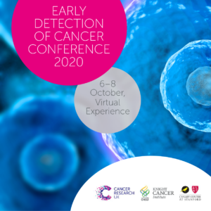
Cancer Research UK, OHSU Knight Cancer Institute and the Canary Center at Stanford, present the Early Detection of Cancer Conference series. The annual Conference brings together experts in early detection from multiple disciplines to share ground breaking research and progress in the field.
The Conference is part of a long-term commitment to invest in early detection research, to understand the biology behind early stage cancers, find new detection and screening methods, and enhance uptake and accuracy of screening.
The 2020 conference will take place October 6-8 virtually.
Cancer Research UK, OHSU Knight Cancer Institute and the Canary Center at Stanford, have been closely monitoring developments relating to the coronavirus (COVID-19) outbreak and reviewing guidance from government bodies. After careful consideration, we have made the decision to convert the Early Detection of Cancer Conference 2020 to a virtual conference, instead of the scheduled in-person conference on October 6-8 in London, UK.
For more information visit the website: http://earlydetectionresearch.com/
CEDSS: “The Origins and Detection of Lethal Prostate Cancer”
Paul Boutros, Ph.D., M.B.A.
Director, Cancer Data Sciences
UCLA
Please see zoom details below:
Meeting URL: https://stanford.zoom.us/s/93515779500
Dial: +1 650 724 9799 or +1 833 302 1536
Meeting ID: 935 1577 9500
Meeting Passcode: 767148
ABOUT
Boutros earned his B.Sc. degree from the University of Waterloo in Chemistry in 2004, and his Ph.D. degree from the University of Toronto, Canada, in Medical Biophysics in 2008. At Toronto, he also earned an executive M.B.A. from the Rothman School of Management. In 2008, Boutros started his independent research career at the Ontario Institute for Cancer Research first as a fellow (2008–2010) and then as principal investigator (2010–2018). He moved to California to join the UCLA faculty in 2018.
Hosted by: Utkan Demirci, Ph.D.
Sponsored by the Canary Center & the Department of Radiology
Stanford University – School of Medicine
PHIND Seminar Series: Serum Modulation of Mitochondrial Function as a Scalable Sensor of Insulin Resistance
Andrew Lipchik, Ph.D.
Postdoctoral Fellow – Michael Snyder Lab
Stanford University
11:00am – 12:00pm Seminar & Discussion
12:00pm – 12:15pm Reception & Light Refreshments
RSVP: https://stanford.zoom.us/webinar/register/7716009863360/WN_dbeuo7csS8q_AhR88XET0g
Location: Zoom
Webinar URL: . https://stanford.zoom.us/s/96358568342
Webinar ID: 963 5856 8342
Dial: +1 650 724 9799 or +1 833 302 1536 (Toll Free)
Password: 767148
ABSTRACT
The global epidemic of obesity is associated with the dramatic increase in the prevalence of type 2 diabetes mellitus (T2D) with an estimated 400 million people worldwide will have T2D by 2030. T2D is proceeded by insulin resistance (IR) for up to decades prior to onset of T2D. Current estimates suggest approximately one in three individuals are sufficiently insulin resistant to be at risk for IR complications including T2D, coronary heart disease and nonalcoholic fatty liver disease. IR often goes undiagnosed due to the complex, invasive and laborious nature of clamp assays preventing their universal application in the clinic. Surrogate measurements using fasting plasma glucose and insulin levels can estimate IR but are imprecise. There is a need for the identification of new biomarkers and assays for the detection and monitoring of IR. Here, we demonstrate the utility of cellular mitochondrial respiration in response to individuals’ serum as a sensor for personalized monitoring of insulin sensitivity. The modulation of insulin-dependent mitochondrial function by patient serum was highly correlated with insulin sensitivity as determined by the gold-standard modified insulin suppression test (IST). We further applied this methodology to monitor insulin sensitivity over time in response to illness as well as treatment with the insulin sensitizing medication, pioglitazone. Our results demonstrate the development and application of a novel surrogate measurement for the determination and monitoring of insulin sensitivity. This assay offers the advantages of minimal invasiveness and complexity compared to IST as well as superior correlation with IST compared to existing surrogate measurements.
ABOUT ANDREW LIPCHIK
Andrew Lipchik majored in Chemistry at Xavier University where he preformed research on the development of oxygen activation Ni(II) complexes with Dr. Craig Davis and Dr. Michael Baldwin at the University of Cincinnati. He went on to obtain his PhD from Purdue University under mentorship of Dr. Laurie Parker. His thesis work focused on identifying determinants of kinase substrate specificity. This understanding was applied to the development of novel kinase-specific peptide biosensors to monitor intracellular kinase activity. Following his graduate work, he joined the laboratory of Michael Snyder at Stanford University where he has focused on understanding the impact of the immune system on insulin resistance and glucose metabolism.
Hosted by: Garry Gold, M.D.
Sponsored by the PHIND Center and the Department of Radiology

ZOOM LINK HERE
“High Resolution Breast Diffusion Weighted Imaging”
Jessica McKay, PhD
ABSTRACT: Diffusion-weighted imaging (DWI) is a quantitative MRI method that measures the apparent diffusion coefficient (ADC) of water molecules, which reflects cell density and serves as an indication of malignancy. Unfortunately, however, the clinical value of DWI is severely limited by the undesirable features in images that common clinical methods produce, including large geometric distortions, ghosting and chemical shift artifacts, and insufficient spatial resolution. Thus, in order to exploit information encoded in diffusion characteristics and fully assess the clinical value of ADC measurements, it is first imperative to achieve technical advancements of DWI.
In this talk, I will largely focus on the background of breast DWI, providing the clinical motivation for this work and explaining the current standard in breast DWI and alternatives proposed throughout the literature. I will also present my PhD dissertation work in which a novel strategy for high resolution breast DWI was developed. The purpose of this work is to improve DWI methods for breast imaging at 3 Tesla to robustly provide diffusion-weighted images and ADC maps with anatomical quality and resolution. This project has two major parts: Nyquist ghost correction and the use of simultaneous multislice imaging (SMS) to achieve high resolution. Exploratory work was completed to characterize the Nyquist ghost in breast DWI, showing that, although the ghost is mostly linear, the three-line navigator is unreliable, especially in the presence of fat. A novel referenceless ghost correction, Ghost/Object minimization was developed that reduced the ghost in standard SE-EPI and advanced SMS. An advanced SMS method with axial reformatting (AR) is presented for high resolution breast DWI. In a reader study, AR-SMS was preferred by three breast radiologists compared to the standard SE-EPI and readout-segmented-EPI.
“Machine-learning Approach to Differentiation of Benign and Malignant Peripheral Nerve Sheath Tumors: A Multicenter Study”
Michael Zhang, MD
ABSTRACT: Clinicoradiologic differentiation between benign and malignant peripheral nerve sheath tumors (PNSTs) is a diagnostic challenge with important management implications. We sought to develop a radiomics classifier based on 900 features extracted from gadolinium-enhanced, T1-weighted MRI, using the Quantitative Imaging Feature Pipeline and the PyRadiomics package. Additional patient-specific clinical variables were recorded. A radiomic signature was derived from least absolute shrinkage and selection operator, followed by gradient boost machine learning. A training and test set were selected randomly in a 70:30 ratio. We further evaluated the performance of radiomics-based classifier models against human readers of varying medical-training backgrounds. Following image pre-processing, 95 malignant and 171 benign PNSTs were available. The final classifier included 21 features and achieved a sensitivity 0.676, specificity 0.882, and area under the curve (AUC) 0.845. Collectively, human readers achieved sensitivity 0.684, specificity 0.742, and AUC 0.704. We concluded that radiomics using routine gadolinium enhanced, T1-weighted MRI sequences and clinical features can aid in the evaluation of PNSTs, particularly by increasing specificity for diagnosing malignancy. Further improvement may be achieved with incorporation of additional imaging sequences.

PHIND Seminar Series: Identifying Microbiome Markers of Progression of Alzheimer’s Disease
Ami Bhatt, M.D., Ph.D.
Assistant Professor of Medicine (Hematology) and of Genetics
Stanford University
Gavin Sherlock, Ph.D.
Associate Professor of Genetics
Stanford University
11:00am – 12:00pm Seminar & Discussion
RSVP: https://stanford.zoom.us/webinar/register/8016040837299/WN_iBOM7R4XQjOPSb20rkUxbw
Location: Zoom Webinar
Webinar URL: https://stanford.zoom.us/s/99730716280
Webinar ID: 997 3071 6280
Dial: +1 650 724 9799 or +1 833 302 1536 (Toll Free)
Password: 767148
ABOUT AMI BHATT
In perpetual awe of how ‘simple’ microbial organisms can perturb complex, multicellular eukaryotic organisms, Ami Bhatt has chosen to dedicate her research program to inspecting, characterizing and dissecting the microbe-human interface. Nowhere is the interaction between hosts and microbes more potentially impactful than in immunocompromised hosts and global settings where infectious and environmental exposures result in drastic and sometimes fatal health consequences.
Ami’s group identifies problems and questions that arise in the course of routine clinical care. Often in collaboration with investigators at Stanford and beyond, the group applies modern genetic, molecular and computational techniques to seek answers to these questions, better understand host-microbe interactions and decipher how perturbation of these interactions may result in human disease phenotypes.
GAVIN SHERLOCK’S RESEARCH INTERESTS
Adaptive Evolution and the Fitness Landscape: When yeast are evolved under various selective pressures in a chemostat, mutations that arise and provide an adaptive advantage will expand within the population. We have pioneered the use of high throughput sequencing to determine the identity of such mutations, as well as to understand the dynamics of the mutations within the populations, and the interactions between the mutations (such as epistasis). Further, we have developed a DNA barcode based lineage tracking system to determine the distribution of fitness effects (DFE) for newly arising beneficial mutations. We have also characterized what we call the genotype-fitness map for beneficial mutations, and have investigated why beneficial mutations provide a positive fitness effect. We are also interested in how beneficial mutations trade-off for different traits, and how those trade-offs constrain adaptive evolution.
Hosted by: Garry Gold, M.D.
Sponsored by the PHIND Center and the Department of Radiology

Ge Wang, PhD
Clark & Crossan Endowed Chair Professor
Director of the Biomedical Imaging Center
Rensselaer Polytechnic Institute
Troy, New York
Abstract:
AI-based tomography is an important application and a new frontier of machine learning. AI, especially deep learning, has been widely used in computer vision and image analysis, which deal with existing images, improve them, and produce features. Since 2016, deep learning techniques are actively researched for tomography in the context of medicine. Tomographic reconstruction produces images of multi-dimensional structures from externally measured “encoded” data in the form of various transforms (integrals, harmonics, and so on). In this presentation, we provide a general background, highlight representative results, and discuss key issues that need to be addressed in this emerging field.
About:
AI-based X-ray Imaging System (AXIS) lab is led by Dr. Ge Wang, affiliated with the Department of Biomedical Engineering at Rensselaer Polytechnic Institute and the Center for Biotechnology and Interdisciplinary Studies in the Biomedical Imaging Center. AXIS lab focuses on innovation and translation of x-ray computed tomography, optical molecular tomography, multi-scale and multi-modality imaging, and AI/machine learning for image reconstruction and analysis, and has been continuously well funded by federal agencies and leading companies. AXIS group collaborates with Stanford, Harvard, Cornell, MSK, UTSW, Yale, GE, Hologic, and others, to develop theories, methods, software, systems, applications, and workflows.