[Menu] [Previous] [Next]
TUTORIAL: Nuclear Physics and Tomography
Positron Emission and Image Reconstruction
Use the "Menu" button to jump to the Let's Play PET Main Menu or click on the Next (right arrowhead) and Previous (left arrowhead) buttons to proceed sequentially through the topics and tutorials. Or, you can return to the Department of Molecular and Medical Pharmacology's Home Page.Contents:
Topics:
Positron Emission
Click on image above to view full-size image.
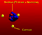
This schematic represents a positron-emitting radioisotope. Positrons (β+) are positively charged electrons. They are emitted from the nucleus of some radioisotopes that are unstable because they have an excessive number of protons and a positive charge. Positron emission stabilizes the nucleus by removing a positive charge through the conversion of a proton into a neutron. In doing this, one element is converted into another, the latter having an atomic number one less than the former. For radioisotopes used in PET, the element formed from positron decay is stable (i.e., not radioactive). All radioisotopes used with PET decay by positron emission. A positron (β+) emitted from a decaying nucleus travels a short distance before colliding with an electron of a nearby atom.
Click on image above to view full-size image.
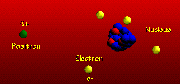
The distance that a positron travels is dependent on its energy.
Click on one or both images above to view full-size images.
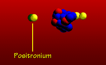
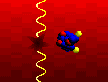
The positron combines with an ordinary electron of a nearby atom in an annihilation reaction, forming positronium as an intermediate.
Click on image above to view full-size image.
When a positron comes in contact with an electron, the two particles annihilate turning the mass of the two particles into two 511-keV gamma-rays that are emitted at 180-degree to each other. These photons easily escape from the human body and can be recorded by external detectors. When detected, the 180-degree emission of two gamma-rays following the disintegration of positronium is called a coincidence line. Coincidence lines provide a unique detection scheme for forming tomographic images with PET.
Emission Detection
Click on image above to view full-size image.
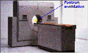
Now let's examine how the tomograph detects the 511-keV gamma rays emitted from the annihilation of positrons with electrons. In a PET study, one administers a positron-emitting radioisotope by injection or inhalation. The isotope then circulates through the bloodstream to reach, for example, brain tissue or cardiac muscle. As positron annihilation occurs, the tomograph detects the isotope's location and concentration.
The line that appears after a positron annihilation event represents the emission of two 511-keV gamma rays at approximately 180-degree apart from one another. The tomograph's job is to detect these coincident rays, which indicates that positron annihilation has occurred somewhere along that coincidence line.
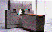
When the 511-keV gamma rays interact with scintillation crystals composed of bismuth germanate (BGO), they are converted into light photons in the crystals.
Click on image above to view full-size image.
Shown here in schematic form, the light photons are converted to electrical signals that are registered by the tomograph's electronics. This conversion and recording process happens almost instantly, so that the scintillation events can be compared among all opposing detectors (along many coincidence lines).
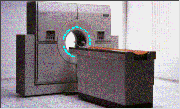
The spatial and temporal distribution of the positron-emitting radioisotope depends on how the organ being scanned handles it biochemically and physiologically. Visualized here are the positron annihilation events and the subsequent gamma-ray emissions.
Having visualized the positron emission detection and recording process, let's look briefly at the image reconstruction process.
Click on image above to view full-size image.
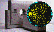
The ring of squares schematically represents one ring of detectors in a PET scanner, which may, for example, have fifteen such rings for simultaneous tomography of many transaxial slices.
Click on image above to view full-size image.
Each detector can be operated in multiple coincidence with many detectors across from it, thereby defining coincidence sampling paths over many angles (fan-beam response).
Click on image above to view full-size image.
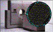
Also, at any given angle many parallel coincidence sampling paths can be defined, resulting in high "linear sampling." These sampling features affect final image quality.
Click on image above to view full-size image.
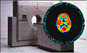
The tomograph's reconstruction software then takes the coincidence events measured at all angular and linear positions to reconstruct an image that depicts the localization and concentration of the positron-emitting radioisotope within a plane of the organ that was scanned.
[Menu] [Previous] [Next]