[Menu] [Previous] [Next]
TUTORIAL: Clinical PET - Oncology
Use the "Menu" button to jump to the Let's Play PET Main Menu or click on the Next and Previous buttons to proceed sequentially through the topics and tutorials. Or, you can return to the Department of Molecular and Medical Pharmacology's Home Page.Contents:
Topics:
Head and Neck
Click on image above to view full-size image.
A person living in the United States has a lifetime chance of one in four of developing cancer and, if inflicted, a 50% chance of dying. Head and neck cancers account for 5% of all cancers. Since advanced lesions have poor survival rates and disproportionate morbidity, the early detection of lesions is especially important. In addition, detection of recurrent lesions, especially following surgery or radiotherapy is critical. Symptoms of head & neck cancer include sore throat, oral bleeding, dysphagia, chronic cough, shortness of breath, hoarseness, unexplained weight loss and nasal obstriction.
Click on image above to view full-size image.
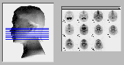
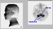
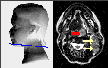
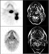
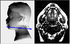
Shown above are a series of FDG-PET scans taken from the upper cervical region of the neck of a patient with a brainstem glioma. The approximate anatomical position for each scan is provided on the model on the left.
Click on image above to view full-size image.
At higher magnification, we can readily see many of the structures of the upper cervical region of the neck. The darker regions reveal structures of high metabolism relative to the lighter regions.
Click on image above to view full-size image.
Click on image above to view full-size image.
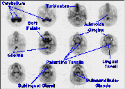
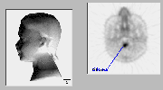
This section, from a more superior aspect of the cervical region reveals the metabolically active cerebellum, part of the central nervous system. The hypermetabolism in the vicinity of the brainstem is due to the presence of a high-grade glioma. Normally, the brainstem and spinal cord are only slightly more active than the surrounding tissue.
Click on image above to view full-size image.
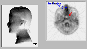
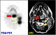
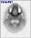
FDG-PET image through the high nasopharynx. The turbinates (nasal conchae) are covered with a metabolically-active mucosa involved in the circulation and filtration of inhaled air. This tissue also contains a vascular space involved in heat exchange. There is also increased activity in the lymphoid tissue and the mucosa of the nasopharynx (red arrow).

Click on image above to view full-size image.
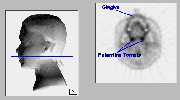
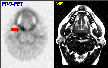
The adenoids (pharyngeal tonsils) are a collection of lymphiod nodules on the posterior wall of the nasopharynx that can only occasionally be differentiated from adjacent structurs using FDG-PET. The presence of a brainstem giloma obstructs the preence of the spinal cord, which would otherwise show a moderate uptake of FDG, similar to the adenoids.
Click on image above to view full-size image.
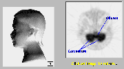
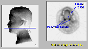
In this plane the hypermetabolic brainstem glioma is can be clearly delineated from adjacent structures.
Click on image above to view full-size image.
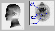
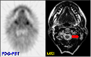
The metabolically active structures in this scan are the soft palate and the parotid glands. The soft palate forms the posterior portion of the roof of the mouth and is lined with mucus membranes. The parotid glands are one of three pairs of salivary glands that secrete their contents into the oral cavity.
Click on image above to view full-size image.
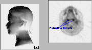
The gingiva (gums) are metabolically slightly more active than surrounding tissue. The palantine tonsils are oval masses of lymphoid tissue located on either side of the back of the tongue and generally show intense activity in FDG-PET scans.
Click on image above to view full-size image.
FDG-PET images through the tongue. The palantine tonsils are oval masses of lymphoid tissue located on either side of the back of the tongue. The lingual tonsil is located at the base of the tongue. The tonsils contain reticuloendothelial cells called macrophages that serve to filter lymph of bacteria and cell debris.
Click on image above to view full-size image.
The palantine tonsils are oval masses of lymphoid tissue located on either side of the back of the tongue. They generally show intense activity in FDG-PET scans.
Click on image above to view full-size image.
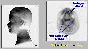
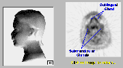
The sublingual and submandibular glands are salivary glands that secrete their contents into the oral cavity. The uptake of FDG in the sublingual gland is generally more intense than that of the submandibular glands (see also plane 11).
Click on image above to view full-size image.
The sublingual and submandibular glands are salivary glands that secrete their contents into the oral cavity.
Case 1
50 year-old female who presented with a left neck mass. Physical examination also revealed a palpable lesion in the left base of the tongue
Click on image above to view full-size image.
Shown above is a pre-treatment MRI image of a 50-year-old female with a poorly differentiated T2N2 squamous cell carcinoma of the base of the tongue. The MRI reveals both the primary tumor and the involvement of two left cervical lymph nodes.
Click on image above to view full-size image.
Now examine the corresponding pre-treatment FDG-PET image for this patient. The intense metabolic activity of the tumor and the involvement of cervical nodes is readily apparent in the FDG-PET image. The extent of the tumor can be gauged by examining the cinematic set of FDG-PET scans on the left, although this would also be apparent in the complete set of MRI images.
Click on image above to view full-size image.
At 6 weeks following radiation treatment, the patient had a persistent 2 cm left neck mass on physical exam, and the MRI scan showed persistent adenopathy (red arrow) and post-radiation changes in the oropharynx with the primary tumor being no longer visible. The FDG-PET scan shows a marked resolution of tumor activity in both the primary tumor and lymph nodes. Fine needle aspirate of the cervical mass was non-diagnostic. This mass gradually decreased in size on physical examination, and the patient has continued to do well clinically.
Click on image above to view full-size image.
Both surgery and radiotherapy cause edema and loss of tissue planes, making detection of lesions challenging using standard imaging techniques such as CT or MRI. In addition, small lymph nodes may contain metastatic foci. FDG-PET imaging is capable of detecting active tumor foci even in small lymph nodes, and is especially helpful in detecting recurrence following therapy. This is particularly critical in cases of head and neck cancer, where one-third of patients die prematurely due to late diagnosis and inadequate treatment of recurrence.
Case 2
58 year-old female with a palpable mass in the base of the tongue. Biopsy revealed squamous cell carcinoma.
Click on image above to view full-size image.
Shown above is a pre-treatment MRI image of a 58 year-old female with a palpable mass in the base of the tongue. The MRI was reported as negative.
Click on image above to view full-size image.
Shown above are pre-treatment FDG-PET images from this patient. Increased metabolic activity is notable on the right side of the tongue (red arrow). Biopsy of the mass revealed squamous cell carcinoma.
Click on image above to view full-size image.
FDG-PET images taken 10 weeks following completion of radiotherapy showed symmetrical FDG uptake with no abnormality in the affected area. The patient has remained clinically free of disease and the mass is not palpable on physical exam. This case demonstrates the sensitivity of FDG-PET in the detection of small, metabolically active tumors and the usefulness of PET in determining resolution of tumor following treatment.
Credits
Material for this section was kindly provided by:Carl Hoh, M.D.
Dept. of Molecular and Medical Pharmacology
UCLA School of Medicine
Yong Choi, Ph.D.
Dept. of Radiology
University of Pittsburgh School of Medicine
Randall Hawkins, M.D., Ph.D.
Nuclear Medicine Division
UCSF School of Medicine
Sheila Rege, M.D.
Dept. of Radiology
LSU Medical School
[Menu] [Previous] [Next]