[Menu] [Previous] [Next]
TUTORIAL: Clinical PET - Oncology
Use the "Menu" button to jump to the Let's Play PET Main Menu or click on the Next and Previous buttons to proceed sequentially through the topics and tutorials. Or, you can return to the Department of Molecular and Medical Pharmacology's Home Page.Contents:
Topics:
Musculoskeletal System
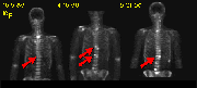
Click on image above to view full-size image.
Whole-body 18F- scans are useful for the detection of bone tumors as well as for studying the progression of these tumors over time. 18F- is particularly suited for the study of bone tumors because it is readily incorporated into bone. Regions displaying increased 18F- incorporation (e.g., arrows) are indicative of tumor formation.
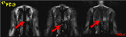
Click on image above to view full-size image.
In addition to 18F- scans, whole-body FDG-PET scans can be used for the initial detection of tumors because of the increased glucose metabolism evident in tumor cells (e.g., arrows). The relatively low uptake of FDG in normal bone is advantageous for identification of metabolically active metastatic bone lesions. Combined FDG-PET and 18F- evaluations of the musculoskeletal system offer the potential of both high sensitivity and high specificity for metastatic and primary bone tumor detection.
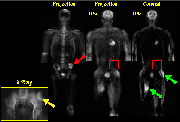
Click on image above to view full-size image.
The above scans are from a patient with treated osteosarcoma. The X Ray reveals a sclerotic lesion in the left iliac wing (yellow arrow). The 18F- shows intense uptake in the left calvarium and iliac wing (red arrow), and right inferior sacral joint, suggesting stimulated osteoblastic activity. However, no corresponding focal increases in FDG metabolism are seen (red boxes). These findings are consistent with treated disease. Note that the asymmetry of FDG uptake in the muscles of the lower extremities (green arrows) is due to the patient's ambulation with a prosthetic limb device. The intense metabolic activity of ambulated skeletal muscle stresses the importance of the patient remaining inactive during the uptake period after FDG injection.
Credits
Material for this section was kindly provided by:Carl Hoh, M.D.
Dept. of Molecular and Medical Pharmacology
UCLA School of Medicine
Yong Choi, Ph.D.
Dept. of Radiology
University of Pittsburgh School of Medicine
Magnus Dahlbom, Ph.D.
Dept. of Molecular and Medical Pharmacology
UCLA School of Medicine
Randall Hawkins, M.D., Ph.D.
Nuclear Medicine Division
UCSF School of Medicine
[Menu] [Previous] [Next]