[Menu] [Previous] [Next]
TUTORIAL: Clinical PET - Oncology
Use the "Menu" button to jump to the Let's Play PET Main Menu or click on the Next and Previous buttons to proceed sequentially through the topics and tutorials. Or, you can return to the Department of Molecular and Medical Pharmacology's Home Page.Contents:
Topics:
Abdomen and Pelvis
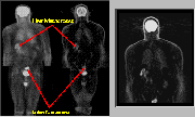
Click on image above to view full-size image.
Evaluation of lesions within or close to the bladder will be hampered by the intense accumulation of urinary FDG activity. Irrigation of the bladder with sterile saline is an option that may be used to decrease the activity in the bladder when there are specific concerns of pelvic or lower abdominal tumors.
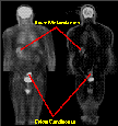
Click on image above to view full-size image.
Depicted above is a patient with metastatic colon carcinoma. Note the proximity of the primary lesion to the bladder. In addition to detecting the primary colon carcinoma, the FDG-PET technique also allows for the preoperative detection of liver metastases. In the above example, liver metastases are readily apparent. Metastases from colon carcinomas often are found in the liver due to the direct drainage of venous blood from the colon into the liver.
Credits
Material for this section was kindly provided by:Carl Hoh, M.D.
Dept. of Radiological Sciences and
Dept. of Molecular and Medical Pharmacology
UCLA School of Medicine
Yong Choi, Ph.D.
Dept. of Radiology
University of Pittsburgh School of Medicine
Randall Hawkins, M.D., Ph.D.
Nuclear Medicine Division
UCSF School of Medicine
[Menu] [Previous] [Next]