[Menu] [Previous] [Next]
TUTORIAL: Clinical PET - Oncology
Use the "Menu" button to jump to the Let's Play PET Main Menu or click on the Next and Previous buttons to proceed sequentially through the topics and tutorials. Or, you can return to the Department of Molecular and Medical Pharmacology's Home Page.Contents:
Topics:
Oncological Scan Evaluation
The use of PET has traditionally been focused on studies of single organ systems such as the brain or heart. With the introduction of PET scanners that allow for whole-body PET in the clinical setting, there has been an increased interest in the application of PET in oncology. In particular, PET is now being applied to the functional evaluation of colorectal, lung, head and neck, and intracranial neoplasms. The application of PET to the study of tumors includes the quantification of tumor perfusion, the evaluation of tumor metabolism, and tracing of radiolabeled cytostatic agents (reviewed in Strauss & Conti, 1991 and Hawkins, et al., 1992).
Click on image above to view full-size image.
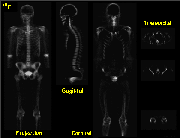
Cancer is characteristically a multisystem disease; however, most of the current PET studies of tumors have been displayed in a transaxial imaging format with the anatomic range of the image set determined by the axial field of view of the given tomograph. A large field of view PET technique, the "whole-body" method, offers the potential to monitor malignant spread of disease in many areas of the body. This whole-body technique is useful for identifying active tumor foci during the initial diagnostic evaluation of a newly diagnosed cancer patient, for monitoring the progression or regression of malignancies, and for monitoring of therapeutic interventions. Two common tracers for whole-body imaging are [18F-] and [18F]FDG. The uptake of F- in the skeleton is proportional to the osteoblastic activity, and the uptake mechanism is an ion exchange between the F- and a hydroxyl ion at the hydroxyapatite crystal surface in the bone. One of the attractive features of F- is the high specific activity of uptake into bone. The uptake of FDG in soft tissue is proportional to glycolysis. The rational for using FDG in tumor localization is that an increase in glycolysis is a biochemical indicator of a malignancy. The above whole-body 18F- images are from a normal patient.
In the standard protocol for whole-body scanning, the patient is injected intravenously with 10mCi of either F- or FDG. The uptake period is 30 min for FDG and 1 hr for F-. The patient is then placed in the scanner and data is acquired over a period varying from 30 to 120 min. The first half of the whole-body image acquisition is initiated from the head down to the waist. The second half of the acquisition requires repositioning the patient to image the lower half of the body. Each half of the whole-body acquisition consists of 16 bed positions, 8 of which are interlaced bed positions required for improved Z axis resolution. Depending on the desired image quality, each bed position can be acquired from 2 to 4 minutes, resulting in a total acquisition time of 64 to 128 minutes, respectively. If kinetic information is desired, a transmission scan ( attenuation correction) is performed at a prescribed bed position, prior to the isotope injection, and a dynamic acquistion is integrated into the 60 minute uptake period.
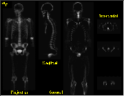
Whole-Body Image Display Formats
Click on image above to view full-size image.
Whole-body image data can be reconstructed and displayed in 2-D projection views, as well as transaxial, coronal and sagittal tomographic views. These images can be displayed individually or in an interactive cine mode, allowing for the analysis of overlying structures.
Click on image above to view full-size image.
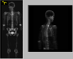
Shown above are 2-D projection images from an 18F- whole-body scan. The image on the left is a shows a normal coronal 18F- projection image. An example of a cinematic image on the right is from a patient with metastatic breast carcinoma. Although less spatially specific, the projection images allow for an overall evaluation of the total-body distribution of 18F- (or FDG) and are thus useful to survey for potential areas of abnormality that later can be fully characterized by tomographic slices. Note that since attenuation correction is typically not applied to the whole-body image data, the reviewer needs to be aware of attenuation artifacts such as the apparent increase in cervical spine activity relative to the thoracic spine due to the patient's arms attenuating the thoracic spine in the lateral projection images.
Click on image above to view full-size image.
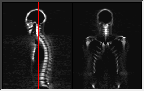
Shown above are 18F- scans (single image; cinematic view) in sagittal and coronal cross-sections. The acquisition time for a whole-body image set is approximately one hour, and proportionally less for more anatomically focused studies. The whole-body PET display formats facilitate comparison with anatomic imaging studies such as X-ray CT and MRI.
Credits
Material for this section was kindly provided by:Carl Hoh, M.D.
Dept. of Radiological Sciences and
Dept. of Molecular and Medical Pharmacology
UCLA School of Medicine
Yong Choi, Ph.D.
Dept. of Radiology
University of Pittsburgh School of Medicine
Magnus Dahlbom, Ph.D.
Dept. of Radiological Sciences and
Dept. of Molecular and Medical Pharmacology
UCLA School of Medicine
Randall Hawkins, M.D., Ph.D.
Nuclear Medicine Division
UCSF School of Medicine
[Menu] [Previous] [Next]