[Menu] [Previous] [Next]
TUTORIAL: Clinical PET - Cardiology
Use the "Menu" button to jump to the Let's Play PET Main Menu or click on the Next and Previous buttons to proceed sequentially through the topics and tutorials. Or, you can return to the Department of Molecular and Medical Pharmacology's Home Page.Contents:
Topics:
Idiopathic Dilated Cardiomyopathy
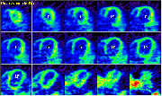
Click on image above to view full-size image.
Shown above are adjacent-plane images from an ammonia study. One can immediately note that the left ventricle (LV) seems larger than in normal studies. Perfusion is relatively homogeneous throughout the myocardium. This condition is referred to as idiopathic dilated cardiomyopathy. Its cause is unknown (idiopathic: arising spontaneously or from an obscure or unknown cause); in contrast to ischemic dilated cardiomyopathy, in which there is an underlying perfusion deficit. PET can be useful in distinguishing idiopathic vs. ischemic dilated cardiomyopathy because flow studies will be normal in the idiopathic case but not in the ischemic case. The dilated cardiomyopathies are characterized by ventricular enlargement with systolic dysfunction eventually leading to congestive heart failure. Ischemic cardiomyopathies can be potentially treated with revascularization, whereas the only treatment for idiopathic dilated cardiomyopathy is cardiac transplantation.
Credits
Material for this section was kindly provided by:Johannes Czernin, M.D.
Dept. of Molecular and Medical Pharmacology
UCLA School of Medicine
Heinrich R. Schelbert, M.D., Ph.D.
Dept. of Molecular and Medical Pharmacology
UCLA School of Medicine
[Menu] [Previous] [Next]