[Menu] [Previous] [Next]
TUTORIAL: Clinical PET - Cardiology
Use the "Menu" button to jump to the Let's Play PET Main Menu or click on the Next and Previous buttons to proceed sequentially through the topics and tutorials. Or, you can return to the Department of Molecular and Medical Pharmacology's Home Page.Contents:
Topics:
Dietary Effects on FDG Metabolism
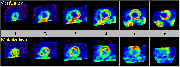
Click on image above to view full-size image.
In general, myocardial utilization of FDG depends on the dietary status of the individual. The normal myocardium uses fatty acids as a primary metabolic fuel. Under conditions of high glucose availability, however,the myocardium does use glucose. Under conditions of low glucose availability, normal tissue relies primarily on fatty acids, whereas abnormal tissue relies on glucose as a fuel for glycolysis. Shown above are studies obtained after having the patient fast for approximately 12 hours. The flow images on the left show decreased perfusion to the anterior and septal regions. The FDG metabolism images show a corresponding hypermetabolism in these same regions. This is due to these hypoperfused areas being primarily dependent on glucose as a metabolic fuel. The other portions of myocardium are adequately perfused and under fasting conditions are primarily dependent on fatty acids.
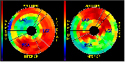
Click on image above to view full-size image.
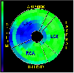
The above represent the flow and metabolism polar maps obtained from the previous images. Note that the flow map shows hypoperfusion in the anterior and septal regions. The metabolism polar map shows increased metabolism (relative to other regions) in the anterior and septal regions.
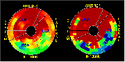
Click on image above to view full-size image.
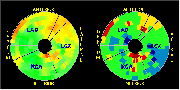
Shown above are the NH3-minus-FDG normalized polar map (left) and the NH3-minus-FDG matched (i.e., normalized to a normal patient data base) polar map (right). The red colors indicate mismatch between NH3 and FDG. Note that there is a definite mismatch in the anterior and septal regions. The severity of this mismatch compared to normals is made very clear in the NH3-FDG matched polar map on the right (very dark red regions).
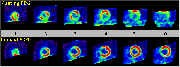
Click on image above to view full-size image.
The same patient was now given glucose, and the FDG uptake study was repeated. Note that in the set of images on the left there is now more homogeneous uptake of FDG by the rest of the myocardium, and the poorly perfused septal/anterior region is relatively hypometabolic. This is also reflected on the polar map on the right. This illustrates that glucose loading allows the normal portions of myocardium to utilize glucose, and these regions actually utilize glucose much better than do the poorly perfused regions. Thus, dietary status significantly affects the FDG metabolic images. Note that FDG studies are normally performed under loaded conditions.
Click on image above to view full-size image.
Shown above are the NH3-minus-FDG normalized polar map (left) and the NH3-minus-FDG matched polar map (right). Note that there is now little mismatch (i.e., few red regions). This study, performed under loaded conditions indicates that viable tissue is present in the antero-septal regions.
Credits
Material for this section was kindly provided by:Johannes Czernin, M.D.
Dept. of Molecular and Medical Pharmacology
UCLA School of Medicine
Heinrich R. Schelbert, M.D., Ph.D.
Dept. of Molecular and Medical Pharmacology
UCLA School of Medicine
[Menu] [Previous] [Next]