![]()
![]()
![]()
![]()
![]()
![]()
![]()
![]()
|
|
|
|
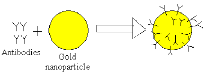
Nanotubes covered in monoclonal antibodies allowed detection of cancer cells. In the experiment, the cancer cells were in a drop of water. Because cancer cells are covered in protein antigens, the antibodies of the nanotubes seeked out the proper antigens on the cancer cells. The specific antibodies used detected insulin-like growth factor 1 receptor (IGF1R). The nanotubes were placed between two electrodes and when cancer cells were binded to them, and an increase in current was measured. The current increase was caused by a rush of electrons from the nanotube device into the cell when they bind. [23] Gold nanoparticles with similar attached antibodies have also been used to identify cancer cells. In another experiment, the protein, epidermal growth factor receptor (EFGR), was used as the target for antibodies attached to the nanoparticles. With darkfield microscopy, cells with attached gold nanoparticles shine while others remain dark. Because healthy cells don’t have EFGR, only cancerous cells are detected. The color of light reflected by the nanoparticles depends on the shape and size of the particle, so adjustments can be made to allow the detection of multiple cells. In other experiments, quantum dots have been used to tag cells. These dots have semiconductor crystals, though, and the effects of them could be toxic. [24]
|
|
Copyright © 2005
Nanogroup Beta: Jason Feng, Maryam Liaqat, Eric Shubo Ma |
Physics 87N: Prof. Hari Manoharan
|