[Menu] [Previous] [Next]
TUTORIAL: Clinical PET - Neurology
Use the "Menu" button to jump to the Let's Play PET Main Menu or click on the Next and Previous buttons to proceed sequentially through the topics and tutorials. Or, you can return to the Department of Molecular and Medical Pharmacology's Home Page.Contents:
Topics:
Parkinson's Disease
Parkinson's disease (idiopathic parkinsonism) is characterized by three major symptoms: rigidity, tremor, and akinesia. Voluntary movements suffer a marked retardation, while muscular strength is well preserved. Speech is often slow and monotonous. Parkinson's Disease is caused by a reduction in dopamine containing nerve cells of the midbrain in the substantia nigra that project to the caudate nucleus (putamen). Once approximately 80% of such cells die, the patient begins to develop symptoms of bradykinesia, immobile facies, stooped posture and resting tremor. Ten to thirty percent of Parkinson's patients also develop dementia. Pathologically there is considerable overlap in the findings of these latter patients with individuals who have pure Alzheimer's disease.
Parkinson's Disease Staging
Parkinson's disease affects 1% of the US population over 50 years of age. The differential diagnosis in patients with suspected Parkinson's disease is very important because therapeutic agents have little or no effect on other Parkinsonian syndromes. Similarly, agents used in conditions that can be mistaken for Parkinson's (e.g. essential tremor) are relatively ineffective for Parkinson's disease. Causes for secondary parkinsonism, including postinfectous, drug-induced, and toxin-induced (MPTP), must also be ruled out. It is also becoming increasingly important to diagnose Parkinson's disease early, as new trends in treatment of this disease are based on early treatment that delay the natural progression of the disease.

Click on image above to view full-size image.
Parkinson's disease is believed to be caused by a reduction in nerve cells in the substantia nigra. These midbrain neurons send projections to the striatum (putamen), where they release the neurotransmitter dopamine. The loss of these cells results in a decreased concentration of endogenous striatal dopamine. Once approximately 80% of these cells die, the patient begins to develop symptoms. The putamen of Parkinson's patients show a characteristic deficit in DOPA uptake. The symptoms of Parkinson's disease can sometimes be alleviated by treatment with L-DOPA, which increases the amount of dopamine that is synthesized in the patient's brain. This is believed to facilitate the activity of the remaining dopaminergic neurons.

Click on image above to view full-size image.
Shown above is the synthetic pathway for dopamine. The first enzyme in this pathway, tyrosine hydroxylase, converts the amino acid tyrosine into L-DOPA. Tyrosine hydroxylase appears to be the rate limiting step in the conversion of tyrosine to catecholamine neurotransmitters. The second enzyme in this pathway is DOPA- decarboxylase. Since this enzyme is not specific to L-DOPA, and can act on all naturally occurring aromatic L-amino acids, it is also referred to as "L-aromatic amino acid decarboxylase." As already mentioned, L-DOPA is often administered to Parkinson's patients because it increases the amount of dopamine that is synthesized in their brain. L-DOPA is often administered in combination with carbidopa to prevent the peripheral conversion of L-DOPA to dopamine (producing a number of undesirable side effects). Dopamine cannot be administered directly because, unlike L-DOPA, dopamine does not cross the blood-brain-barrier. Most patients starting therapy require 300-1000 mg of levodopa per day for adequate response. A patient must have their individual levels titrated with respect to improvement of symptoms and development of side effects. Side effects include dystonia, confusion, hallucinations, dizziness, hypotension, and autonomic dysfunction. Perhaps the most disturbing side effect is an "on-off" phenomenon in which the patients symptoms actually worsen with therapy.
The deficit in dopamine uptake characteristic of Parkinson's disease can be readily seen using radiolabeled L-DOPA and positron emission tomography. In addition FDG-PET studies of glucose metabolism can be used to differentiate the dementia that often accompanies Parkinson's disease from the movement deficits that are associated with pathology of the basal ganglia.

Click on image above to view full-size image.
These scans are from a normal patient. The two FDG-PET scans show normal metabolism throughout the cortex, while the corresponding F-DOPA scan shows normal DOPA uptake in the striatum.

Click on image above to view full-size image.
This FDG-PET scan of a Parkinson's patient without dementia shows normal glucose metabolism.

Click on image above to view full-size image.
However, the corresponding F-DOPA scan reveals the decreased dopamine synthesis that is characteristic of Parkinson's disease.

Click on image above to view full-size image.
This F-DOPA scan, from another Parkinson's patient, shows a similar pattern of low dopamine synthesis.

Click on image above to view full-size image.
The corresponding FDG scan of this other Parkinson's patient reveals a pattern of cortical hypometabolism that is characteristic of dementia (examine the cortex adjacent to the dotted lines). Thus, by using both F-DOPA and FDG, PET provides the opportunity to identify the source of dementia independent of the diagnosis of Parkinson's disease.
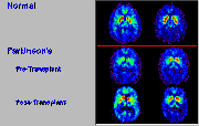
Carbidopa-levodopa therapy has markedly improved the life expectancy and quality of life of Parkinson's patients; unfortunately patients eventually continue to deteriorate and alternative solutions have to be explored. Clinical trials in patients with Parkinson's disease suggest that the grafting of dopamine- producing cells derived from human fetal tissue may be beneficial for alleviating many of the symptoms associated with Parkinson's disease, and for reducing by up to 60% the dose of L-DOPA required by these patients.

Click on image above to view full-size image.
Compare the 18F-DOPA scan of the patient with Parkinson's disease with that of the normal volunteer. Note the reduced uptake of DOPA in the basal ganglia in the Parkinson's patient. Following surgical transplantation of fetal dopamine synthesizing cells into the striatum, uptake of DOPA in the in the Parkinson's patient shows dramatic improvement.
Credits
Material for this section was kindly provided by:
Michael E. Phelps, Ph.D.
Dept. of Molecular and Medical Pharmacology
UCLA School of Medicine
John Mazziotta, M.D., Ph.D.
Dept. of Molecular and Medical Pharmacology and
Dept. of Neurology
UCLA School of Medicine
[Menu] [Previous] [Next]